Line Drawing 1

Fig. 12 Rhinebothrium fungiforme n. sp. from Hemitrygon fluviorum. A, Bothridial surface pattern in glycerine mount; B, Scolex; C,
Entire cestode; D, Premature segments; E, Mature terminal segment. S... MoreFig. 12 Rhinebothrium fungiforme n. sp. from Hemitrygon fluviorum. A, Bothridial surface pattern in glycerine mount; B, Scolex; C,
Entire cestode; D, Premature segments; E, Mature terminal segment. Scale-bars: A, D, E, 100 µm; B, 200 µm; C. 1.0 mm. |
Line Drawing 2
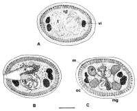
Fig. 13 Rhinebothrium fungiforme n. sp. from Hemitrygon fluviorum. Transverse histological sections through mature segment. A,
Section through preporal region of segment showing testes and vitelline ... MoreFig. 13 Rhinebothrium fungiforme n. sp. from Hemitrygon fluviorum. Transverse histological sections through mature segment. A,
Section through preporal region of segment showing testes and vitelline follicles; B, Section through cirrus-sac and terminal vagina; C,
Section through ovarian isthmus. Scale-bar: 50 µm. Abbreviations: cs, cirrus-sac; m, muscle bundle; mg, Mehlis gland; oc,
osmoregulatory canal; t, testis; v, vagina; vi, vitelline follicle. |
Photo Micrograph
|
Scanning Electron Micrograph

Fig. 14 Rhinebothrium fungiforme n. sp. from Hemitrygon fluviorum. Scanning electron micrographs. A, Scolex; B, Filiform
microtriches on distal surface of bothridium; C, Gladiate spinitriches of pedi... MoreFig. 14 Rhinebothrium fungiforme n. sp. from Hemitrygon fluviorum. Scanning electron micrographs. A, Scolex; B, Filiform
microtriches on distal surface of bothridium; C, Gladiate spinitriches of pedicels and cephalic peduncle; D, Microtriches at junction of
cephalic peduncle and strobila. Scale-bars: A, 10 µm; BD, 1 µm. |
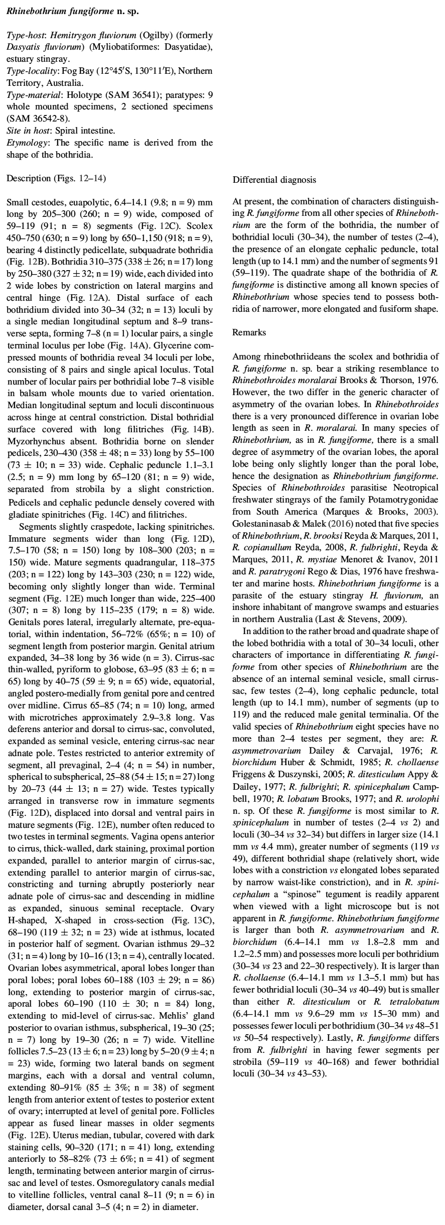
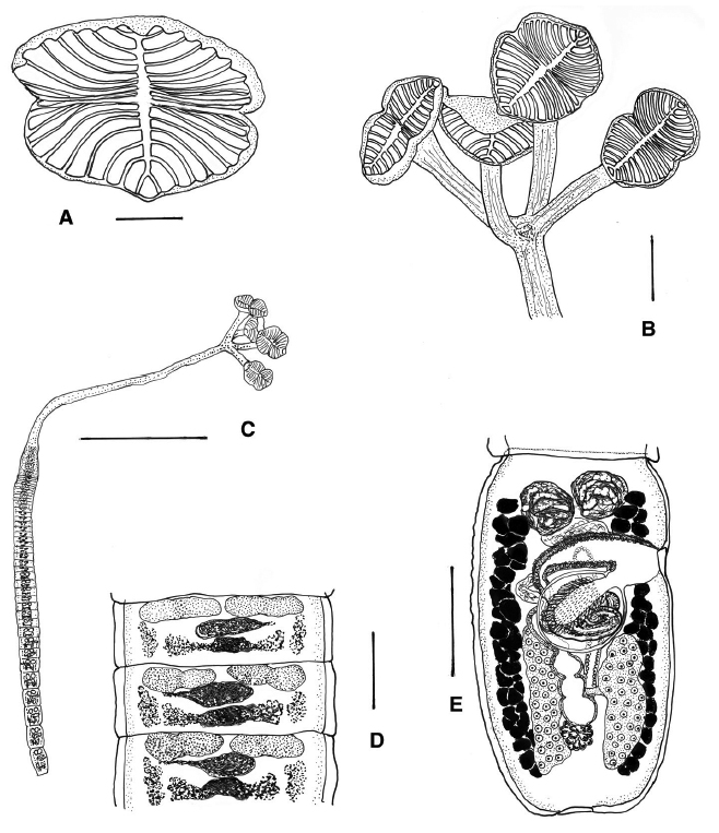 Fig. 12 Rhinebothrium fungiforme n. sp. from Hemitrygon fluviorum. A, Bothridial surface pattern in glycerine mount; B, Scolex; C,
Entire cestode; D, Premature segments; E, Mature terminal segment. Scale-bars: A, D, E, 100 µm; B, 200 µm; C. 1.0 mm.
Fig. 12 Rhinebothrium fungiforme n. sp. from Hemitrygon fluviorum. A, Bothridial surface pattern in glycerine mount; B, Scolex; C,
Entire cestode; D, Premature segments; E, Mature terminal segment. Scale-bars: A, D, E, 100 µm; B, 200 µm; C. 1.0 mm.  Fig. 13 Rhinebothrium fungiforme n. sp. from Hemitrygon fluviorum. Transverse histological sections through mature segment. A,
Section through preporal region of segment showing testes and vitelline follicles; B, Section through cirrus-sac and terminal vagina; C,
Section through ovarian isthmus. Scale-bar: 50 µm. Abbreviations: cs, cirrus-sac; m, muscle bundle; mg, Mehlis gland; oc,
osmoregulatory canal; t, testis; v, vagina; vi, vitelline follicle.
Fig. 13 Rhinebothrium fungiforme n. sp. from Hemitrygon fluviorum. Transverse histological sections through mature segment. A,
Section through preporal region of segment showing testes and vitelline follicles; B, Section through cirrus-sac and terminal vagina; C,
Section through ovarian isthmus. Scale-bar: 50 µm. Abbreviations: cs, cirrus-sac; m, muscle bundle; mg, Mehlis gland; oc,
osmoregulatory canal; t, testis; v, vagina; vi, vitelline follicle. 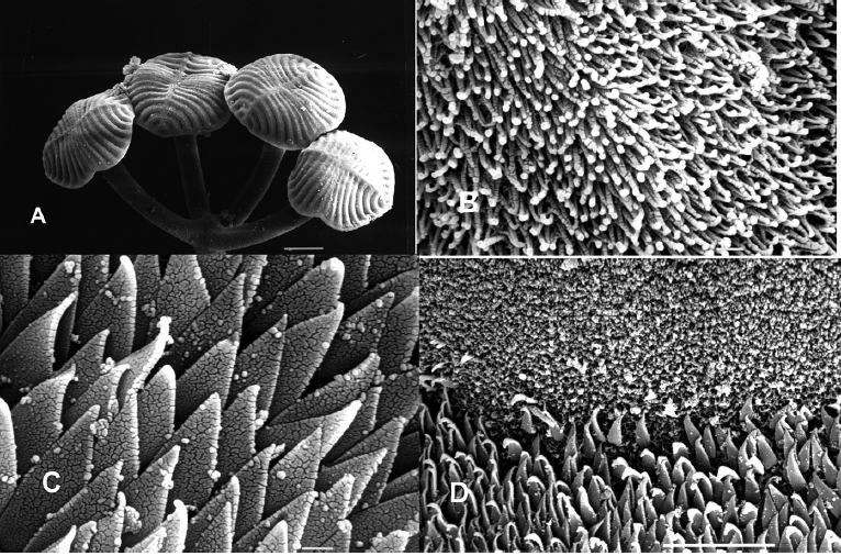 Fig. 14 Rhinebothrium fungiforme n. sp. from Hemitrygon fluviorum. Scanning electron micrographs. A, Scolex; B, Filiform
microtriches on distal surface of bothridium; C, Gladiate spinitriches of pedicels and cephalic peduncle; D, Microtriches at junction of
cephalic peduncle and strobila. Scale-bars: A, 10 µm; BD, 1 µm.
Fig. 14 Rhinebothrium fungiforme n. sp. from Hemitrygon fluviorum. Scanning electron micrographs. A, Scolex; B, Filiform
microtriches on distal surface of bothridium; C, Gladiate spinitriches of pedicels and cephalic peduncle; D, Microtriches at junction of
cephalic peduncle and strobila. Scale-bars: A, 10 µm; BD, 1 µm. 

