Cestode Scientific Name
| Species ID | 14053 |
|---|---|
| Order | Rhinebothriidea |
| Family | Anthocephaliidae |
| Subfamily | |
| Genus | Anthocephalum |
| Species | jeancadenati |
| Authority | Boudaya, Neifar, & Euzet, 2018 |
| Taxonomic Status | Valid |
| Valid Name | |
| Synonyms | |
| Genus Record | No |
| Type Species | No |
| Verified | No |
| Verified By | |
| Citation(s) |
Boudaya, L., L. Neifar, and L. Euzet. 2018. A new genus and three new species of Anthocephaliidae (Cestoda, Rhinebothriidea) from the round fantail stingray, Taeniurops grabata (Chondrichthyes, Dasyatidae) from the Mediterranean Sea and Atlantic Ocean. Journal of Helminthology : 111. (7129) Download PDF |
| Redescription | |
| Scientific Name Notes |
Record Data
| Date (MM/DD/YYYY) | Action | User Name |
|---|---|---|
| 06/14/2019 | Created | K. Herzog |
| 06/14/2019 | Modified | K. Herzog |
Type Host
| Host Class | Elasmobranchii | ||||||
|---|---|---|---|---|---|---|---|
| Host Order | Myliobatiformes | ||||||
| Host Family | Dasyatidae | ||||||
|
Type Host (Literal) |
|
||||||
|
Type Host (Valid) |
|
||||||
| Additional Host(s) | |||||||
| Site in Host | spiral intestine | ||||||
| Host Notes | "Prevalence in Tunisia: 100% (16 of 16 specimens examined)." |
Type Locality
| Country | Tunisia |
|---|---|
| Body of Water | |
| Island(s) | |
| City/Region | Zarzis |
| Coordinates | 33°15'N, 11°10'E |
| DD Latitude | |
| DD Longitude | |
| Additional Localities | Bizerte (37°30'N, 9°50'E), Tunisia; Sfax (34°47'N, 10°51'E), Tunisia; Gorée Island (14°40'N, 18°25'W), Senegal. |
| Locality Notes |
Specimens
| Type Material | holotype (MNHN HEL831); 10 paratypes (MNHN HEL832839). |
|---|---|
| Total Number of Type Specimens | 11 |
| Voucher Material | |
| Specimen Notes |
Data are given as in original description unless otherwise indicated.
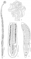
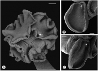
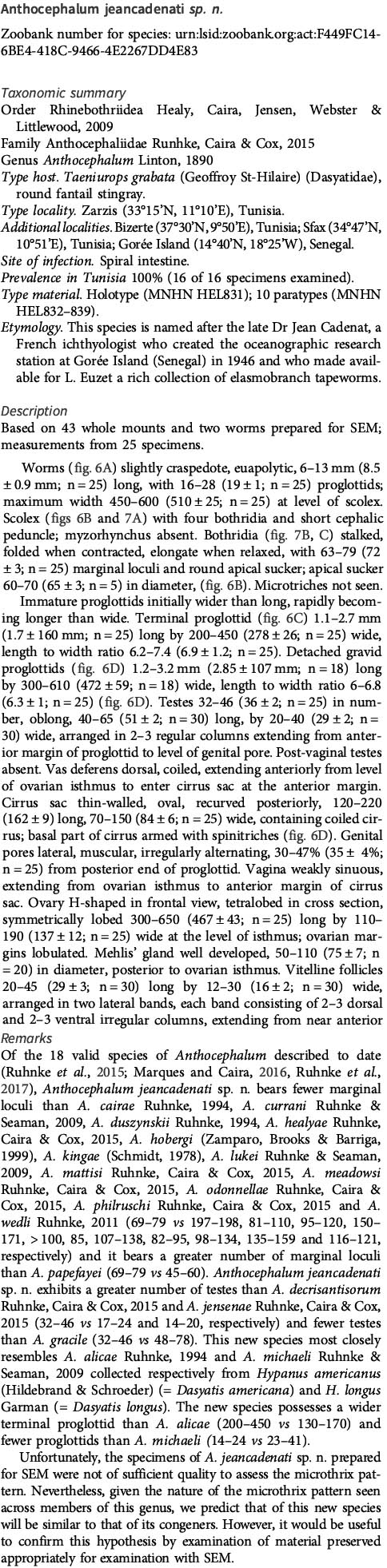
 Fig. 6. Anthocephalum jeancadenati sp. n. (A) Entire worm; (B) scolex, apical view; (C) mature proglottid, ventral view; (D) gravid proglottid, ventral view. Scale bars: A = 500 µm; B, C, D = 250 µm.
 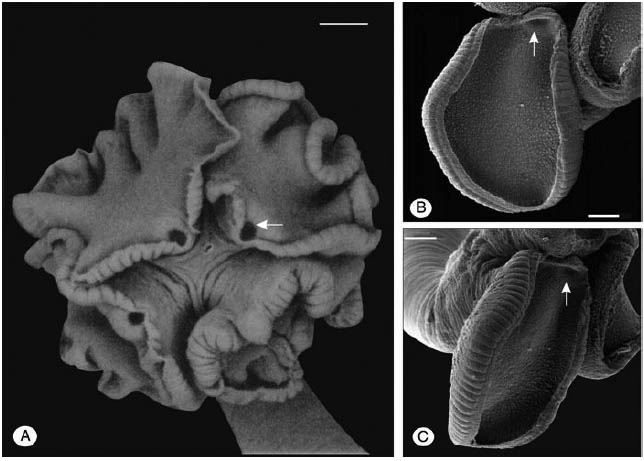 Fig. 7. Scanning electron micrographs of Anthocephalum jeancadenati sp. n. (A) Scolex in apical view, showing apical sucker; (b) distal bothridial surface; (C) bothridia, showing marginal loculi. Arrows indicate the apical sucker. Scale bars: A = 100 µm; B = 50 µm.
 Best viewed in Firefox


