Line Drawing 1
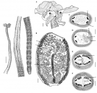
Fig. 1 Orygmatobothrium persiense n. sp. (entire worm). Scale bar = 1 mm. Fig. 2 Orygmatobothrium persiense
n. sp. a scolex, b mature
proglottid, c cross section of
mature proglottid at level of te... MoreFig. 1 Orygmatobothrium persiense n. sp. (entire worm). Scale bar = 1 mm. Fig. 2 Orygmatobothrium persiense
n. sp. a scolex, b mature
proglottid, c cross section of
mature proglottid at level of testes
anterior to cirrus sac, d cross
section of mature proglottid at
level of cirrus sac, e cross section
of mature proglottid at level
of uterus and vagina anterior to
ovarian isthmus, f cross section
of mature proglottid at level of
Mehlis gland and ovary, Abbreviations:
c cirrus, cs cirrus
sac, oc osmoregulatory canal,
ov ovary, mg Mehlis gland, t
testis, u uterus, v vagina, vd vas
deferens, vi vitelline follicle.
Scale bars: af = 100 μm. |
Line Drawing 2
|
Photo Micrograph
|
Scanning Electron Micrograph

Fig. 3 Surface ultrastructure of Orygmatobothrium persiense n. sp. a
scolex, b apical sucker, c acicular filitriches on outer surface of apical
sucker in vicinity of distal bothridial surface, d cen... MoreFig. 3 Surface ultrastructure of Orygmatobothrium persiense n. sp. a
scolex, b apical sucker, c acicular filitriches on outer surface of apical
sucker in vicinity of distal bothridial surface, d central accessory
organ, e acicular filitriches on central accessory organ, f, g gongylate
columnar spinitriches on distal bothridial surface; inset in F shows
enlarged detail of microtriches, h acicular filitriches on marginal part
of distal bothridial surface, i trifid spinitriches on proximal bothridial
surface, arrowheads indicate lateral prongs, j cephalic peduncle and
anterior part of germinative zone, k aristate gladiate spinitriches on
cephalic peduncle, arrowheads indicate apical projection (aristate) of
spinithrix, l scutes on proglottid, m densely packed acicular filitriches
forming scutes. Scale bars: a = 500 μm, b, d = 20 μm, c, e, h, i, k,
m = 1 μm, f, g = 2 μm, j, l = 50 μm. |
 Fig. 1 Orygmatobothrium persiense n. sp. (entire worm). Scale bar = 1 mm. Fig. 2 Orygmatobothrium persiense
n. sp. a scolex, b mature
proglottid, c cross section of
mature proglottid at level of testes
anterior to cirrus sac, d cross
section of mature proglottid at
level of cirrus sac, e cross section
of mature proglottid at level
of uterus and vagina anterior to
ovarian isthmus, f cross section
of mature proglottid at level of
Mehlis gland and ovary, Abbreviations:
c cirrus, cs cirrus
sac, oc osmoregulatory canal,
ov ovary, mg Mehlis gland, t
testis, u uterus, v vagina, vd vas
deferens, vi vitelline follicle.
Scale bars: af = 100 μm.
Fig. 1 Orygmatobothrium persiense n. sp. (entire worm). Scale bar = 1 mm. Fig. 2 Orygmatobothrium persiense
n. sp. a scolex, b mature
proglottid, c cross section of
mature proglottid at level of testes
anterior to cirrus sac, d cross
section of mature proglottid at
level of cirrus sac, e cross section
of mature proglottid at level
of uterus and vagina anterior to
ovarian isthmus, f cross section
of mature proglottid at level of
Mehlis gland and ovary, Abbreviations:
c cirrus, cs cirrus
sac, oc osmoregulatory canal,
ov ovary, mg Mehlis gland, t
testis, u uterus, v vagina, vd vas
deferens, vi vitelline follicle.
Scale bars: af = 100 μm. 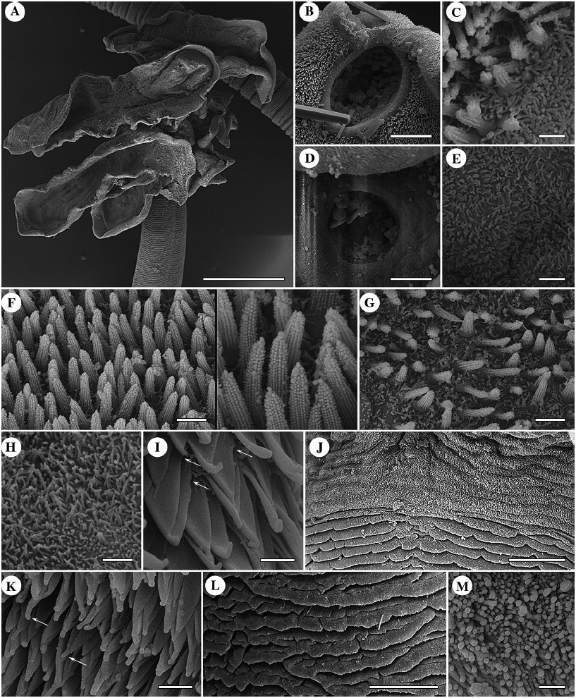 Fig. 3 Surface ultrastructure of Orygmatobothrium persiense n. sp. a
scolex, b apical sucker, c acicular filitriches on outer surface of apical
sucker in vicinity of distal bothridial surface, d central accessory
organ, e acicular filitriches on central accessory organ, f, g gongylate
columnar spinitriches on distal bothridial surface; inset in F shows
enlarged detail of microtriches, h acicular filitriches on marginal part
of distal bothridial surface, i trifid spinitriches on proximal bothridial
surface, arrowheads indicate lateral prongs, j cephalic peduncle and
anterior part of germinative zone, k aristate gladiate spinitriches on
cephalic peduncle, arrowheads indicate apical projection (aristate) of
spinithrix, l scutes on proglottid, m densely packed acicular filitriches
forming scutes. Scale bars: a = 500 μm, b, d = 20 μm, c, e, h, i, k,
m = 1 μm, f, g = 2 μm, j, l = 50 μm.
Fig. 3 Surface ultrastructure of Orygmatobothrium persiense n. sp. a
scolex, b apical sucker, c acicular filitriches on outer surface of apical
sucker in vicinity of distal bothridial surface, d central accessory
organ, e acicular filitriches on central accessory organ, f, g gongylate
columnar spinitriches on distal bothridial surface; inset in F shows
enlarged detail of microtriches, h acicular filitriches on marginal part
of distal bothridial surface, i trifid spinitriches on proximal bothridial
surface, arrowheads indicate lateral prongs, j cephalic peduncle and
anterior part of germinative zone, k aristate gladiate spinitriches on
cephalic peduncle, arrowheads indicate apical projection (aristate) of
spinithrix, l scutes on proglottid, m densely packed acicular filitriches
forming scutes. Scale bars: a = 500 μm, b, d = 20 μm, c, e, h, i, k,
m = 1 μm, f, g = 2 μm, j, l = 50 μm. 

