Cestode Scientific Name
| Species ID | 14044 |
|---|---|
| Order | Onchoproteocephalidea II |
| Family | |
| Subfamily | |
| Genus | Acanthobothrium |
| Species | marquesi |
| Authority | Rodríguez-Ibarra, Pulido-Flores, Violante-González & Monks, 2018 |
| Taxonomic Status | Valid |
| Valid Name | |
| Synonyms | |
| Genus Record | No |
| Type Species | No |
| Verified | No |
| Verified By | |
| Citation(s) |
Rodríguez-Ibarra, E., G. Pulido-Flores, J. Violante-González, and S. Monks. 2018. A new species of Acanthobothrium (Eucestoda: Onchobothriidae) in Aetobatus cf. narinari (Myliobatidae) from Campeche, México. Brazilian Journal of Veterinary Parasitology 27(1): 6774. (7127) Download PDF |
| Redescription | |
| Scientific Name Notes | Zoobank: B4DAA0D5-284B-4B66- 9477‑CDDEAA98B7BE |
Record Data
| Date (MM/DD/YYYY) | Action | User Name |
|---|---|---|
| 06/11/2019 | Created | K. Herzog |
| 06/11/2019 | Modified | K. Herzog |
Type Host
| Host Class | Elasmobranchii | ||||||
|---|---|---|---|---|---|---|---|
| Host Order | Myliobatiformes | ||||||
| Host Family | Aetobatidae | ||||||
|
Type Host (Literal) |
|
||||||
|
Type Host (Valid) |
|
||||||
| Additional Host(s) | |||||||
| Site in Host | spiral intestine | ||||||
| Host Notes |
Type Locality
| Country | México |
|---|---|
| Body of Water | Gulf of Mexico, Laguna de Términos |
| Island(s) | |
| City/Region | Ciudad del Carmen, Campeche |
| Coordinates | 18°3519N, 91°3330W |
| DD Latitude | |
| DD Longitude | |
| Additional Localities | Champotón, Campeche, México (19°21N; 90°54W) |
| Locality Notes |
Specimens
| Type Material | holotype (CNHE 10554); 2 paratypes (CNHE 1055510556); 8 paratypes (HWML 139377139384); 3 paratypes (CHE P00061P00063) |
|---|---|
| Total Number of Type Specimens | 14 |
| Voucher Material | |
| Specimen Notes |
Data are given as in original description unless otherwise indicated.

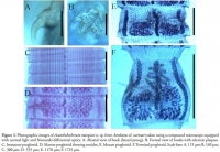

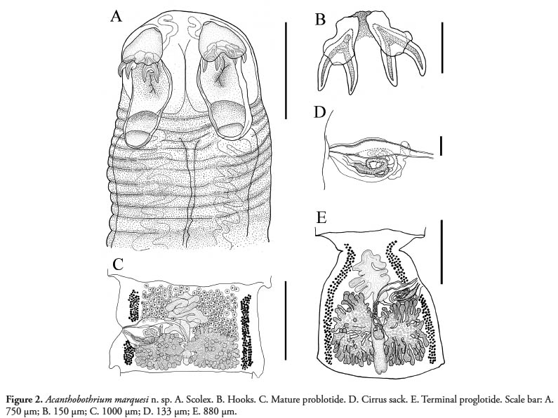 Figure 2. Acanthobothrium marquesi n. sp. A. Scolex. B. Hooks. C. Mature problotide. D. Cirrus sack. E. Terminal proglotide. Scale bar: A. 750 μm; B. 150 μm; C. 1000 μm; D. 133 μm; E. 880 μm.
  Figure 3. Photographic images of Acanthobothrium marquesi n. sp. from Aetobatus cf. narinari taken using a compound microscope equipped with normal light and Nomarski differential optics. A. Abaxial view of hook (lateral prong). B. Formal view of hooks with sclerotic plaques. C. Immature proglottid. D. Mature proglottid showing testicles. E. Mature proglottid. F. Terminal proglottid. Scale bars: A. 115 μm; B. 160 μm; C. 300 μm; D. 535 μm; E. 1170 μm; F. 1725 μm.
 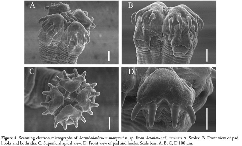 Figure 4. Scanning electron micrographs of Acanthobothrium marquesi n. sp. from Aetobatus cf. narinari A. Scolex. B. Front view of pad, hooks and bothridia. C. Superficial apical view. D. Front view of pad and hooks. Scale bars: A, B, C, D 100 μm.
 Best viewed in Firefox


