Cestode Scientific Name
| Species ID | 14034 |
|---|---|
| Order | Onchoproteocephalidea II |
| Family | |
| Subfamily | |
| Genus | Acanthobothrium |
| Species | omanense |
| Authority | Maleki, Malek, & Palm, 2019 |
| Taxonomic Status | Valid |
| Valid Name | |
| Synonyms | |
| Genus Record | No |
| Type Species | No |
| Verified | No |
| Verified By | |
| Citation(s) |
Maleki, L., M. Malek, and H. W. Palm. 2019. Five new species of Acanthobothrium (Cestoda: Onchoproteocephalidea) from the long-tailed butterfly ray, Gymnura cf. poecilura (Elasmobranchii: Gymnuridae), from the Persian Gulf and Gulf of Oman. Zootaxa 4609(2): 289-307. (7123) Download PDF |
| Redescription | |
| Scientific Name Notes | urn:lsid:zoobank.org:act:741B8217-BB7B-454CAF35-DDAC48E3E694 |
Record Data
| Date (MM/DD/YYYY) | Action | User Name |
|---|---|---|
| 06/10/2019 | Created | K. Jensen |
| 06/10/2019 | Modified | K. Jensen |
| 06/11/2019 | Modified | K. Jensen |
Type Host
| Host Class | Elasmobranchii | ||||||
|---|---|---|---|---|---|---|---|
| Host Order | Myliobatiformes | ||||||
| Host Family | Gymnuridae | ||||||
|
Type Host (Literal) |
|
||||||
|
Type Host (Valid) |
|
||||||
| Additional Host(s) | |||||||
| Site in Host | spiral intestine | ||||||
| Host Notes |
Type Locality
| Country | Iran |
|---|---|
| Body of Water | Gulf of Oman |
| Island(s) | |
| City/Region | |
| Coordinates | 25°11'N, 60°33'E |
| DD Latitude | |
| DD Longitude | |
| Additional Localities | Persian Gulf |
| Locality Notes | Locality given in paper as: 25°11'N, 60°33'E25°25'N, 57°43'E |
Specimens
| Type Material | Holotype (ZCUOK. 117), 5 Paratypes (ZCUOK 118ZCUOK 122), 5 paratypes (ZUTC Platy. 1330ZUTC Platy. 1334), 1 SEM voucher (ZUTC Platy. 1335). |
|---|---|
| Total Number of Type Specimens | 11 mature worms, 1 scolex examined with SEM, and a whole mount of its voucher |
| Voucher Material | |
| Specimen Notes |
Data are given as in original description unless otherwise indicated.

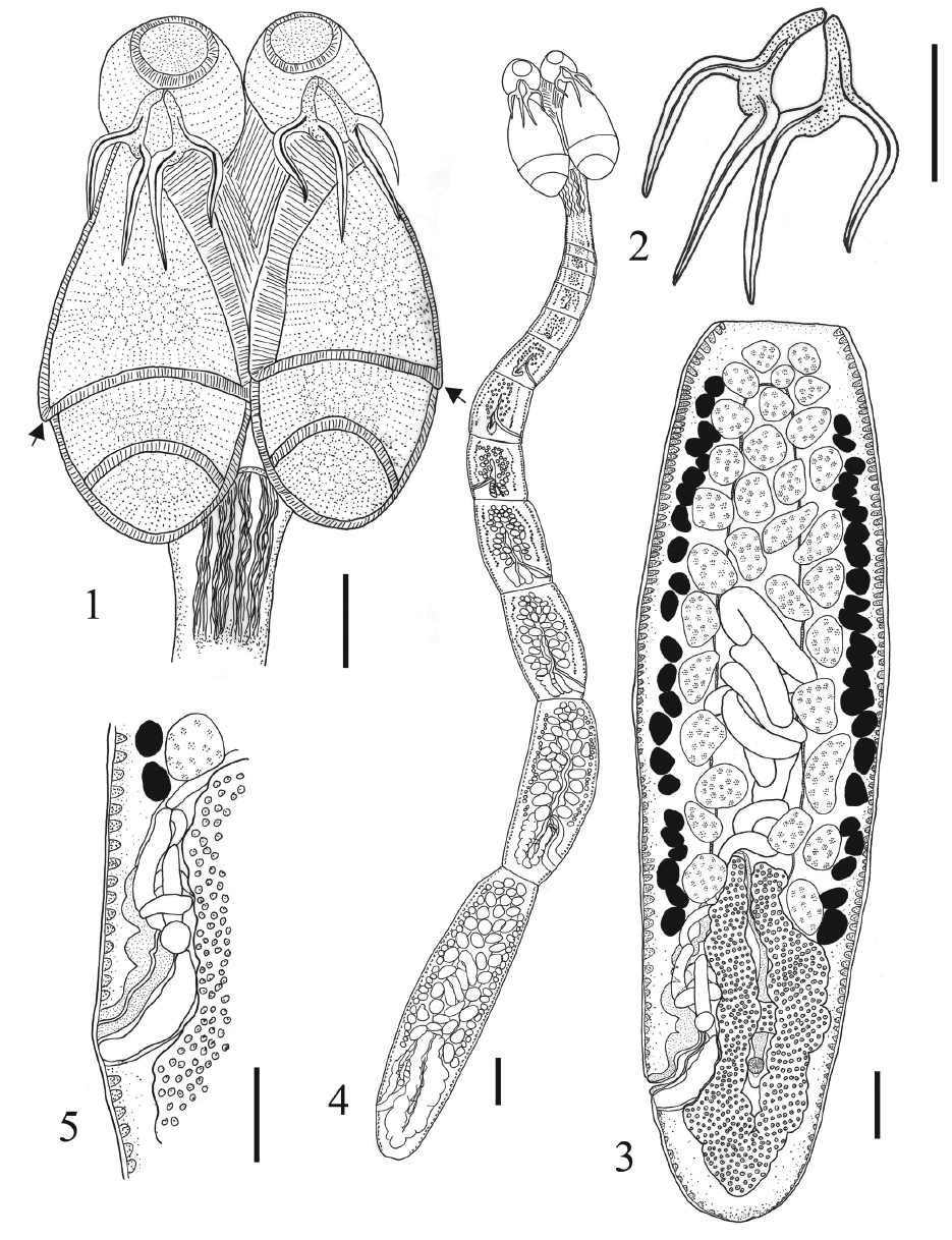 FIGURES 15. Acanthobothrium omanense n. sp. 1. Scolex. Arrows show the marginal lappets. 2. Hooks. 3. Terminal mature proglottid. 4. Whole worm. 5. Terminal genitalia. Scale bars: Figs. 1, 2, 3, 5= 50 μm; Fig. 4= 100 μm.
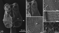
 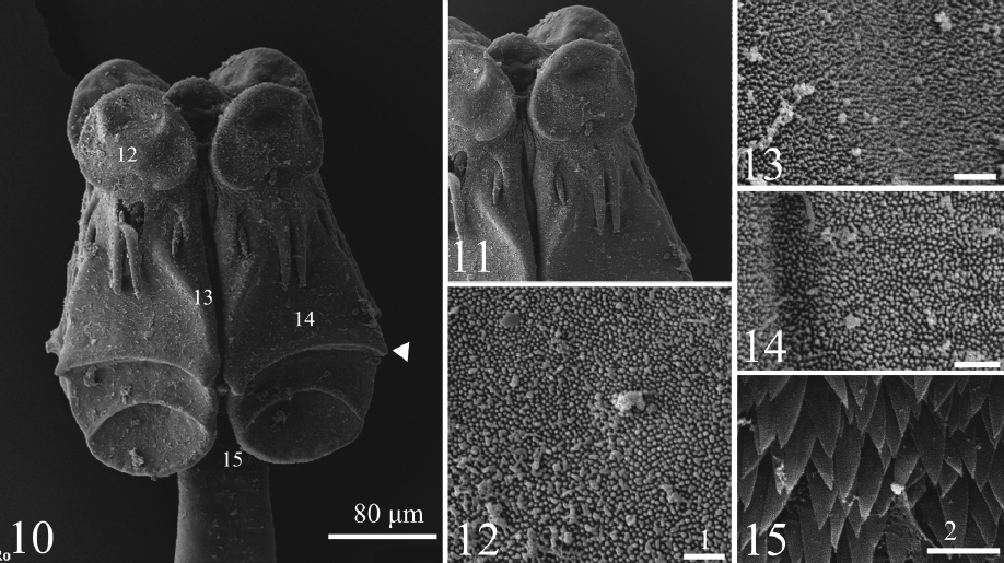 FIGURES 1015. Scanning electron micrographs of Acanthobothrium omanense n. sp. 10. Scolex. Note:
Small numbers on the scolex correspond to the figures showing higher magnification images of these surfaces, and white arrow
tip show the marginal lappet. 11. Apical pad and hooks. 12. Surface of apical pad. 13. Proximal bothridial surface. 14. Distal
bothridial surface. 15. Cephalic peduncle surface. Scale bars: Fig. 10=80 μm; Figs. 11=40 μm; Figs 15=2 μm; Figs. 12, 13, 14=1 μm.
FIGURES 1015. Scanning electron micrographs of Acanthobothrium omanense n. sp. 10. Scolex. Note:
Small numbers on the scolex correspond to the figures showing higher magnification images of these surfaces, and white arrow
tip show the marginal lappet. 11. Apical pad and hooks. 12. Surface of apical pad. 13. Proximal bothridial surface. 14. Distal
bothridial surface. 15. Cephalic peduncle surface. Scale bars: Fig. 10=80 μm; Figs. 11=40 μm; Figs 15=2 μm; Figs. 12, 13, 14=1 μm. Best viewed in Firefox


