Cestode Scientific Name
| Species ID | 14014 |
|---|---|
| Order | Onchoproteocephalidea I |
| Family | Proteocephalidae |
| Subfamily | |
| Genus | Harriscolex |
| Species | piramutab |
| Authority | (Woodland, 1933) de Chambrier, Kuchta & Scholz, 2015 |
| Taxonomic Status | Valid |
| Valid Name | |
| Synonyms | Anthobothrium piramutab Woodland, 1933; Proteocephalus piramutab (Woodland, 1933) Rego, 1984 |
| Genus Record | No |
| Type Species | No |
| Verified | No |
| Verified By | |
| Citation(s) |
Woodland, W. N. F. 1933. On two new cestodes from the Amazon siluroid fish Brachyplatystoma vaillanti Cuv. and Val.. Parasitology 25: 485-490. (3834) Download PDFde Chambrier, A., R. Kuchta, and T. Scholz. 2015. Tapeworms (Cestoda: Proteocephalidea) of teleost fishes from the Amazon River in Peru: additional records as an evidence of unexplored species diversity. Revue Suisse de Zoologie 122(1): 149-163. (6617) Download PDF |
| Redescription | |
| Scientific Name Notes |
Record Data
| Date (MM/DD/YYYY) | Action | User Name |
|---|---|---|
| 12/06/2018 | Created | P. Alves |
| 12/06/2018 | Modified | P. Alves |
| 02/03/2020 | Modified | B. Barbeau |
| 12/03/2021 | Modified | B. Barbeau |
Type Host
| Host Class | |||||||
|---|---|---|---|---|---|---|---|
| Host Order | Siluriformes | ||||||
| Host Family | Pimelodidae | ||||||
|
Type Host (Literal) |
|
||||||
|
Type Host (Valid) |
|
||||||
| Additional Host(s) | |||||||
| Site in Host | anterior intestine | ||||||
| Host Notes |
Type Locality
| Country | Brazil |
|---|---|
| Body of Water | Amazon River |
| Island(s) | |
| City/Region | between Codajás and Parintins, between Codajás and Parintins, Amazon State |
| Coordinates | |
| DD Latitude | |
| DD Longitude | |
| Additional Localities | |
| Locality Notes |
Specimens
| Type Material | |
|---|---|
| Total Number of Type Specimens | |
| Voucher Material | |
| Specimen Notes |
Data are given as in original description unless otherwise indicated.

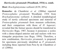
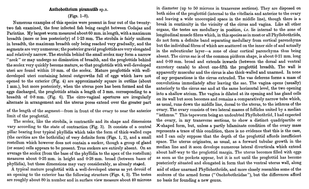
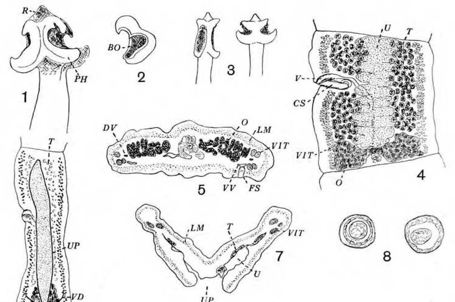 Plate XXX. Anthobothrium piramutab sp.n. (Figs. 1-8). Fig. 1 (x56): the scolex. Fig. 2 (x56): a phyllidium showing the shape of the cavity (bothridium). Fig. 3: diagram showing different forms of the scolex. Fig. 4 (x39): a young gravid proglottid. Fig. 5 (x56): transverse section of young gravid proglottid through hind end of ovary. Fig. 6 (x18): posterior gravid proglottid which has shed its eggs through the elongated uterus pore. Fig. 7 (x56): transverse section of posterior gravid proglottid to show the uterus pore and degeneration of other organs. Fig. 8 (x395): eggs drawn in the original formalin-glycerine preserving fluid.
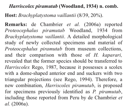
Best viewed in Firefox


