Cestode Scientific Name
| Species ID | 13955 |
|---|---|
| Order | Cyclophyllidea |
| Family | Nematotaeniidae |
| Subfamily | |
| Genus | Lanfrediella |
| Species | amphicirrus |
| Authority | Melo, Giese, Furtado, Soares, Goncalves, Vallinoto & Santos, 2011 |
| Taxonomic Status | Valid |
| Valid Name | |
| Synonyms | |
| Genus Record | No |
| Type Species | Yes |
| Verified | No |
| Verified By | |
| Citation(s) |
Melo, F. T., E. G. Giese, A. P. Furtado, M. J. Soares, E. C. Gonçalves, A. C. Vallinoto and J. N. Santos. 2011. Lanfrediella amphicirrus gen. nov. sp. nov. Nematotaeniidae (Cestoda: Cyclophyllidea), a tapeworm parasite of Rhinella marina (Linnaeus, 1758) (Amphibia: Bufonidae). Memórias do Instituto Oswaldo Cruz 106(6): 670-677. (6823) |
| Redescription | |
| Scientific Name Notes |
Record Data
| Date (MM/DD/YYYY) | Action | User Name |
|---|---|---|
| 03/15/2018 | Created | R. Kuchta |
| 03/15/2018 | Modified | R. Kuchta |
| 12/15/2021 | Modified | R. Kuchta |
Type Host
| Host Class | Amphibia | ||||||
|---|---|---|---|---|---|---|---|
| Host Order | Anura | ||||||
| Host Family | Bufonidae | ||||||
|
Type Host (Literal) |
|
||||||
|
Type Host (Valid) |
|
||||||
| Additional Host(s) | |||||||
| Site in Host | small intestine | ||||||
| Host Notes |
Type Locality
| Country | Brazil |
|---|---|
| Body of Water | |
| Island(s) | |
| City/Region | Belém, state of Pará |
| Coordinates | |
| DD Latitude | |
| DD Longitude | |
| Additional Localities | |
| Locality Notes |
Specimens
| Type Material | -CHIOC 37314a (holotype); CHIOC 37314b, 35706 (paratypes) |
|---|---|
| Total Number of Type Specimens | |
| Voucher Material | |
| Specimen Notes | -the helminthological collection of the Oswaldo Cruz Institute (CHIOC) of the Oswaldo Cruz Foundation in Rio de Janeiro, Brazil |
Data are given as in original description unless otherwise indicated.
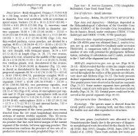

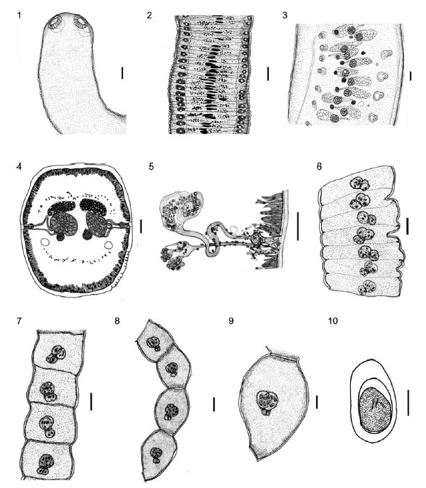 Figs 1-10: light microscopy of Lanfrediella amphicirrus gen. nov. sp. nov. 1: dorsoventral view of the scolex (Bar = 100 μm); 2: immature segments in a dorsoventral view (Bar = 50 μm); 3: mature segments (Bar = 50 μm); 4: cross section of mature segment showing two groups of reproductive organs and two cirrus pouches (Bar = 50 μm); 5: reconstruction of a cirrus pouch with cirri showing the genital atrium, deferent channel and vaginal channel (Bar = 50 μm); 6: pregravid strobila segments showing the initial region of segmentation (Bar = 100 μm); 7: gravid segments showing the maturation of the paruterine capsules and visible segmentation (Bar = 100 μm); 8: posterior extremity showing gravid segments (Bar = 100 μm); 9: detail of a gravid segment where it is possible to observe two paruterine capsules per segment (Bar = 50 μm); 10: details of eggs showing the outer envelope, embriophore, and the oncosphere (Bar = 30 μm).
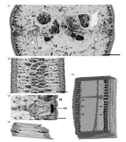
 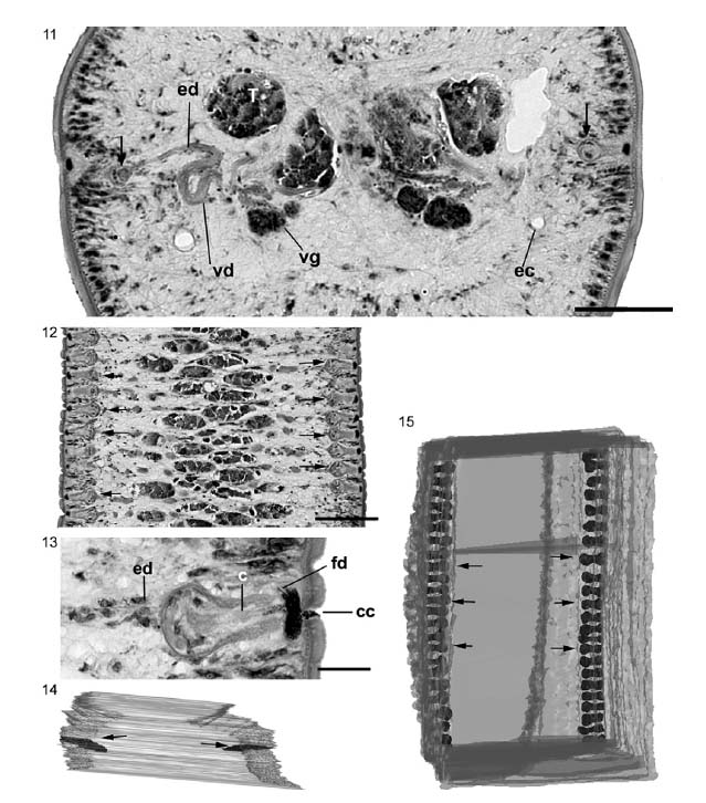 Figs 11-15: longitudinal and cross-sections of mature segments embedded in Historesin and three-dimensional (3D) reconstruction based on serial longitudinal sections of a strobila segment. 11: cross-section of mature segments showing testis (T), ejaculatory ducts (ed), vitelline glands (vg), viteline duct (vd), excretory channels (ec) and two cirrus (c) pouches (arrows) (Bar = 100 μm); 12: longitudinal section of a strobila with mature segments showing the presence of two c pouches (arrows) in each lateral side of the strobila (Bar = 100 μm); 13: detail of a c pouch in a longitudinal section of mature segments in which it is possible to observe the insertion of ed, c, female duct (fd) and the copulatory channel (cc) (Bar = 100 μm); 14: superior view of in the transversal axis of 3D reconstruction where is possible to observe the internal position of two lateral rows of c pouches (arrows); 15: dorsoventral view in 3D reconstruction showing the position of c pouches (arrows) in each lateral margin of the strobila.
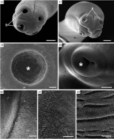
  Figs 16-22: scanning electron microscopy of Lanfrediella amphicirrus gen. nov. sp. nov. 16: general view of relaxed scolex showing the suckers (S), absence of a rosteolum or apical organ (Bar = 100 μm); 17: overall view of contracted scolex showing morphology of S in this moment (Bar = 100 μm); 18: internal details of S (asterisk) in contracted scolex. Note the absence of spines or any other structures (Bar = 10 μm); 19: internal details of S (asterisk) in contracted scolex (Bar = 100 μm); 20: details of the microtriches close to border of S (Bar = 10 μm); 21: details of microtriches in immature segments (Bar = 10 μm); 22: details of microtriches in mature segments (Bar = 10 μm).
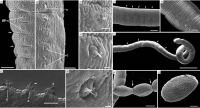
 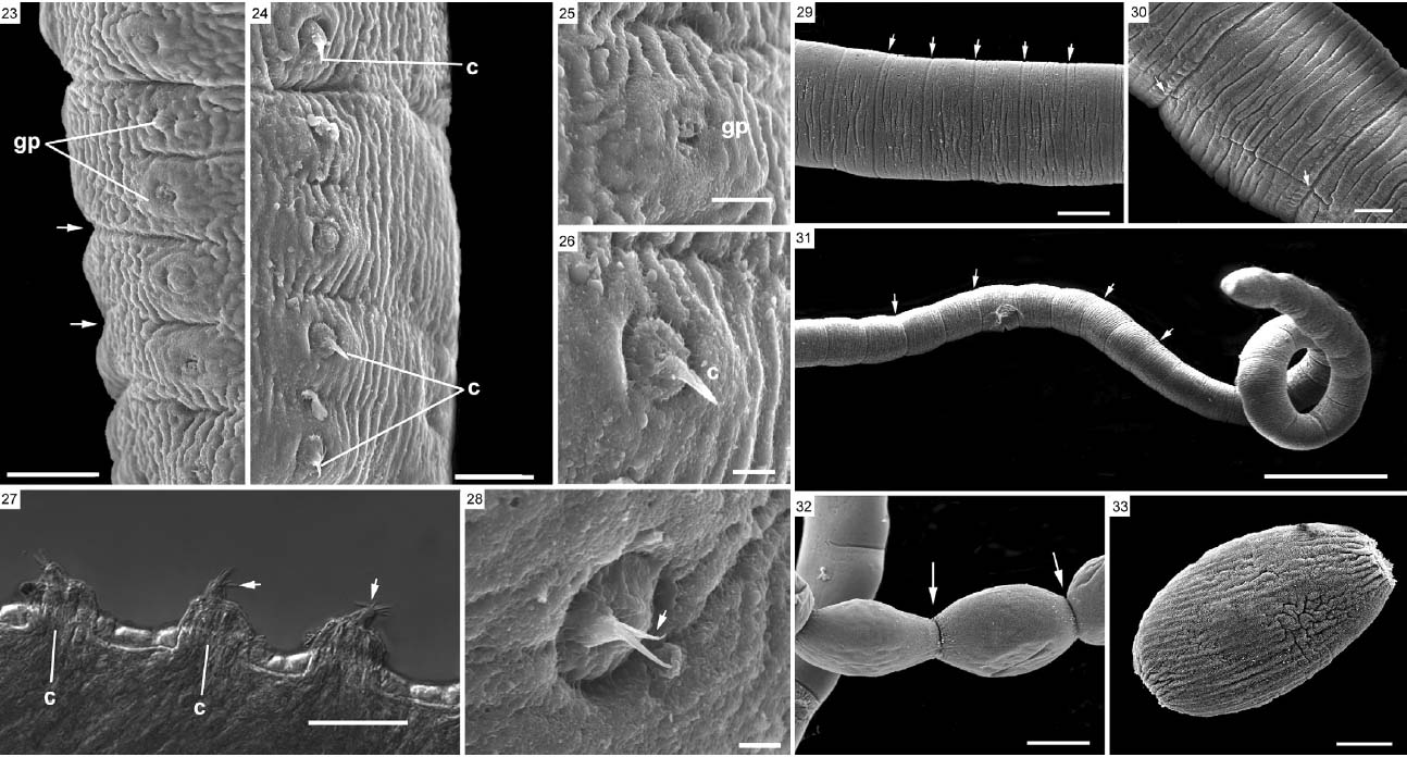 Figs 23-28: scanning electron microscopy of mature segments and differential interference contrast (DIC) of mature segments of Lanfrediella amphicirrus gen. nov. sp. nov. 23: lateral view of a mature segment showing the segmentation (arrow) and genital pores (gp) (Bar = 26 μm); 24: view of the opposite side of the segment in Fig. 23 showing the gp and a partly extroverted (C) (Bar = 20 μm); 25: view of the region of the gp region observed in Fig. 23 (Bar = 10 μm); 26: details of the cirrus (c) located in the mature segment observed in Fig. 24 (Bar = 5 μm); 27: image of DIC of mature segments with the best-adjusted focus obtained using the CombineZP software showing c extroverted with many spines (arrows)
(Bar = 100 μm); 28: partially extroverted c (Bar = 3 μm)
Figs 29-33: scanning electron microscopy of mature and gravid segments of Lanfrediella amphicirrus gen. nov. sp. nov. 29: mature segments of parasite showing the segmentation (arrows) (Bar = 100 μm); 30: gravid segment view in strobila where is possible to observe the segmentation (arrows) (Bar = 100 μm); 31: posterior end of a strobila showing the segmentation of gravid segments (arrows) (Bar = 400 μm); 32: end portion of the parasite showing the constrictions between gravid segments (arrows) (Bar = 100 μm); 33: isolated gravid segment. Note the elliptical morphology, characteristic of this stage, and the absence of genital pores (Bar = 50 μm).
Figs 23-28: scanning electron microscopy of mature segments and differential interference contrast (DIC) of mature segments of Lanfrediella amphicirrus gen. nov. sp. nov. 23: lateral view of a mature segment showing the segmentation (arrow) and genital pores (gp) (Bar = 26 μm); 24: view of the opposite side of the segment in Fig. 23 showing the gp and a partly extroverted (C) (Bar = 20 μm); 25: view of the region of the gp region observed in Fig. 23 (Bar = 10 μm); 26: details of the cirrus (c) located in the mature segment observed in Fig. 24 (Bar = 5 μm); 27: image of DIC of mature segments with the best-adjusted focus obtained using the CombineZP software showing c extroverted with many spines (arrows)
(Bar = 100 μm); 28: partially extroverted c (Bar = 3 μm)
Figs 29-33: scanning electron microscopy of mature and gravid segments of Lanfrediella amphicirrus gen. nov. sp. nov. 29: mature segments of parasite showing the segmentation (arrows) (Bar = 100 μm); 30: gravid segment view in strobila where is possible to observe the segmentation (arrows) (Bar = 100 μm); 31: posterior end of a strobila showing the segmentation of gravid segments (arrows) (Bar = 400 μm); 32: end portion of the parasite showing the constrictions between gravid segments (arrows) (Bar = 100 μm); 33: isolated gravid segment. Note the elliptical morphology, characteristic of this stage, and the absence of genital pores (Bar = 50 μm). Best viewed in Firefox


