Cestode Scientific Name
| Species ID | 13929 |
|---|---|
| Order | Bothriocephalidea |
| Family | Echinophallidae |
| Subfamily | |
| Genus | Acanthocephallus |
| Species | wageneri |
| Authority | (Monticelli, 1890) Lühe, 1910 |
| Taxonomic Status | Synonym |
| Valid Name | Echinophallus wageneri (Monticelli, 1890) Schumacher, 1914 |
| Synonyms | |
| Genus Record | No |
| Type Species | Yes |
| Verified | No |
| Verified By | |
| Citation(s) |
Monticelli, F. S. 1890. Note elmintologiche. Bollettino della Societa dei Naturalisti in Napoli 4(2): 189-208. (4193) Download PDF |
| Redescription | |
| Scientific Name Notes | -preoccupied |
Record Data
| Date (MM/DD/YYYY) | Action | User Name |
|---|---|---|
| 09/18/2017 | Created | R. Kuchta |
| 09/18/2017 | Modified | R. Kuchta |
Type Host
| Host Class | Actinopterygii | ||||||
|---|---|---|---|---|---|---|---|
| Host Order | Percifomes | ||||||
| Host Family | Centrolophidae | ||||||
|
Type Host (Literal) |
|
||||||
|
Type Host (Valid) |
|
||||||
| Additional Host(s) | |||||||
| Site in Host | intestine | ||||||
| Host Notes |
Type Locality
| Country | Italy |
|---|---|
| Body of Water | Mediterranean Sea |
| Island(s) | |
| City/Region | Ligurian Sea off Genoa |
| Coordinates | |
| DD Latitude | |
| DD Longitude | |
| Additional Localities | |
| Locality Notes |
Specimens
| Type Material | -not located |
|---|---|
| Total Number of Type Specimens | |
| Voucher Material | |
| Specimen Notes |
Data are given as in original description unless otherwise indicated.


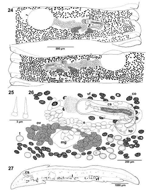 Figs. 2427. Echinophallus wageneri from Outer Hebrides. Fig. 24. Gravid segment; dorsal view; eggs in uterine sac not illustrated. Fig. 25. Detail of cirrus spines. Fig. 26. Terminal genitalia; ventral view. Fig. 27. Cross-section of segment at the level of cirrus-sac. Abbreviations: co corpuscles; cs cirrus-sac; e eggs; mg Mehlis gland; ov ovary; sp sphincter; t testes; vf vitelline follicles.
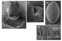
 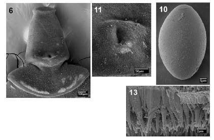 Figs. 6, 10, 11, 13. Echinophallus wageneri from Outer Hebrides. Fig. 6. Scolex in dorsoventral view. Fig. 10. Egg. Fig. 11. Detailed view of posterior margin of bothria. Fig. 13. Detailed view of posterior margin of segment.
 Best viewed in Firefox


