Line Drawing 1
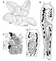
FIGURE 6. Line drawings of Rhinebothrium reydai n. sp. A. Scolex (MIUP CR1, Holotype), B. Terminal, mature proglottid in which testes are atrophied (MZUSP 7931b, Paratype), C. Cirrus-sac (MZUSP 7931p,... MoreFIGURE 6. Line drawings of Rhinebothrium reydai n. sp. A. Scolex (MIUP CR1, Holotype), B. Terminal, mature proglottid in which testes are atrophied (MZUSP 7931b, Paratype), C. Cirrus-sac (MZUSP 7931p, Paratype), D. Subterminal, mature proglottid (MZUSP 7931b, Paratype). |
Line Drawing 2
|
Photo Micrograph
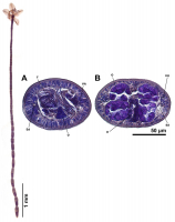
FIGURE 5. Light micrograph of the holotype of Rhinebothrium reydai n. sp. from Styracura schmardae (MIUP CR1). FIGURE 8. Micrographs of transversal histological sections of Rhinebothrium reydai n. sp.... MoreFIGURE 5. Light micrograph of the holotype of Rhinebothrium reydai n. sp. from Styracura schmardae (MIUP CR1). FIGURE 8. Micrographs of transversal histological sections of Rhinebothrium reydai n. sp. (MZUSP 7933a7933d, Paratypes). A. Section at level of testes, B. Section at level of ovary. Abbreviations: Ed. Excretory duct, O. Ovary, T. Testis, U. Uterus, Vd. Vas deferens, Vit. Vitelline follicle. |
Scanning Electron Micrograph
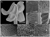
FIGURE 7. Scanning electron micrographs of Rhinebothrium reydai n. sp. (MZUSP 7932, Paratype). A. Scolex, B. Proximal surface of anterior loculus, C. Proximal surface near centre of bothridium, D. Dis... MoreFIGURE 7. Scanning electron micrographs of Rhinebothrium reydai n. sp. (MZUSP 7932, Paratype). A. Scolex, B. Proximal surface of anterior loculus, C. Proximal surface near centre of bothridium, D. Distal surface near centre of bothridium, E. Distal surface of transverse septa, F. Distal surface near longitudinal septum in anterior region of bothridium, G. Proximal surface in posterior region of bothridium, H. Surface of anterior portion of strobila. |

 FIGURE 6. Line drawings of Rhinebothrium reydai n. sp. A. Scolex (MIUP CR1, Holotype), B. Terminal, mature proglottid in which testes are atrophied (MZUSP 7931b, Paratype), C. Cirrus-sac (MZUSP 7931p, Paratype), D. Subterminal, mature proglottid (MZUSP 7931b, Paratype).
  FIGURE 5. Light micrograph of the holotype of Rhinebothrium reydai n. sp. from Styracura schmardae (MIUP CR1). FIGURE 8. Micrographs of transversal histological sections of Rhinebothrium reydai n. sp. (MZUSP 7933a7933d, Paratypes). A. Section at level of testes, B. Section at level of ovary. Abbreviations: Ed. Excretory duct, O. Ovary, T. Testis, U. Uterus, Vd. Vas deferens, Vit. Vitelline follicle.
  FIGURE 7. Scanning electron micrographs of Rhinebothrium reydai n. sp. (MZUSP 7932, Paratype). A. Scolex, B. Proximal surface of anterior loculus, C. Proximal surface near centre of bothridium, D. Distal surface near centre of bothridium, E. Distal surface of transverse septa, F. Distal surface near longitudinal septum in anterior region of bothridium, G. Proximal surface in posterior region of bothridium, H. Surface of anterior portion of strobila.

