Line Drawing 1

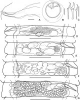
Armadolepis (Armadolepis) tenorai n. sp. A holotype, dorsoventral view of scolex; B paratype (MHNG-PLAT-91137) sublateral view of scolex; C paratype (AM12-50#3), rostellar hooks in profile and ... MoreArmadolepis (Armadolepis) tenorai n. sp. A holotype, dorsoventral view of scolex; B paratype (MHNG-PLAT-91137) sublateral view of scolex; C paratype (AM12-50#3), rostellar hooks in profile and view from anterior surface, showing flattened hook guard; D holotype, male mature proglottides, dorsal view; E holotype, hermaphroditic mature proglottides, dorsal view; F holotype, genital ducts, dorsal view. Scale bars: A, B, F = 100 μm; C = 10 μm; E, F = 300 μm |
Line Drawing 2
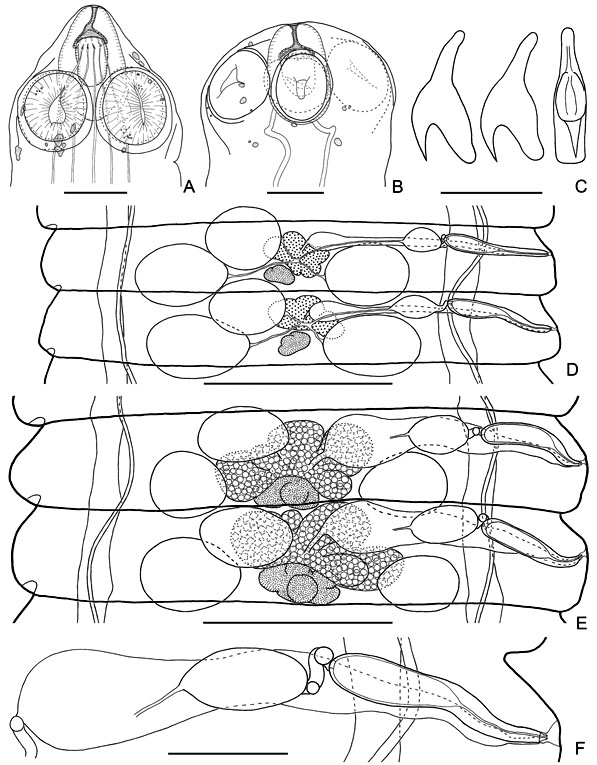
Armadolepis (Armadolepis) tenorai n. sp. A holotype, cirrus and vagina, ventral view; B paratype (AM12-50#3), egg; C paratype (AM12-50#3), embryonic hooks; D paratype (AM12-50#3), first posmat... MoreArmadolepis (Armadolepis) tenorai n. sp. A holotype, cirrus and vagina, ventral view; B paratype (AM12-50#3), egg; C paratype (AM12-50#3), embryonic hooks; D paratype (AM12-50#3), first posmature proglottis from ventral side, showing uterus at early stages; E holotype, terminal posmature proglottis from dorsal side, showing uterine development; F holotype, pregravid proglottis from dorsal side, showing appearance of uterine pockets and perforations; G holotype, gravid proglottis from dorsal side, showing labyrinthine uterus with lateral uterine pockets. Scale bars: A, B = 20 μm; C = 10 μm; D-G = 300 μm |
Photo Micrograph

Armadolepis (Armadolepis) tenorai n. sp. A paratype (AM12-50#3), crown of rostellar hooks; B holotype, dorsoventral view of scolex, showing calcareous corpuscles; C paratype (AM12-50#3) calcareo... MoreArmadolepis (Armadolepis) tenorai n. sp. A paratype (AM12-50#3), crown of rostellar hooks; B holotype, dorsoventral view of scolex, showing calcareous corpuscles; C paratype (AM12-50#3) calcareous corpuscles; D paratype (AM12-50#3), tiny canals in scolex; E paratype (AM12-50#3), egg. Scale bars: A, C, D, E = 20 μm; B = 100 μm |
Scanning Electron Micrograph
|
 Armadolepis (Armadolepis) tenorai n. sp. A holotype, dorsoventral view of scolex; B paratype (MHNG-PLAT-91137) sublateral view of scolex; C paratype (AM12-50#3), rostellar hooks in profile and view from anterior surface, showing flattened hook guard; D holotype, male mature proglottides, dorsal view; E holotype, hermaphroditic mature proglottides, dorsal view; F holotype, genital ducts, dorsal view. Scale bars: A, B, F = 100 μm; C = 10 μm; E, F = 300 μm
 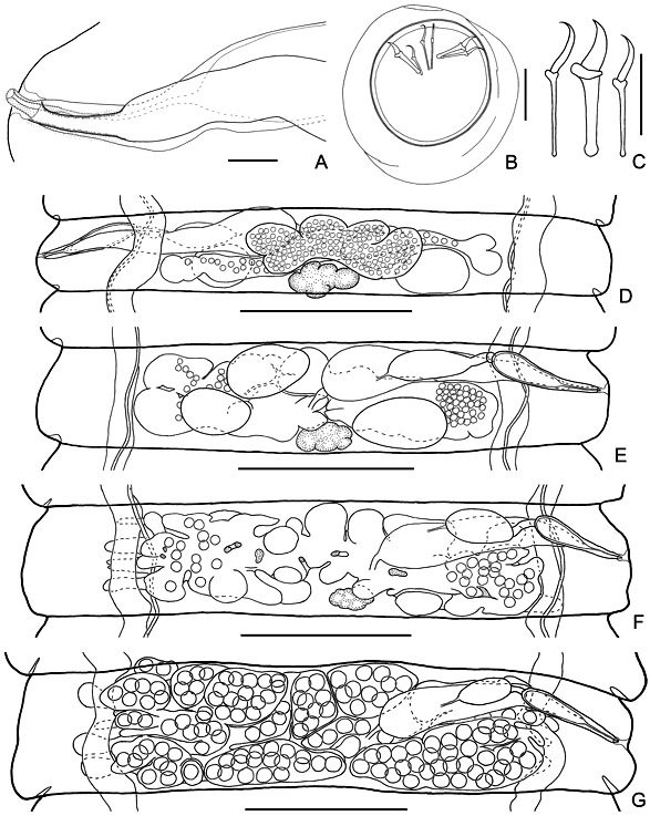 Armadolepis (Armadolepis) tenorai n. sp. A holotype, cirrus and vagina, ventral view; B paratype (AM12-50#3), egg; C paratype (AM12-50#3), embryonic hooks; D paratype (AM12-50#3), first posmature proglottis from ventral side, showing uterus at early stages; E holotype, terminal posmature proglottis from dorsal side, showing uterine development; F holotype, pregravid proglottis from dorsal side, showing appearance of uterine pockets and perforations; G holotype, gravid proglottis from dorsal side, showing labyrinthine uterus with lateral uterine pockets. Scale bars: A, B = 20 μm; C = 10 μm; D-G = 300 μm
  Armadolepis (Armadolepis) tenorai n. sp. A paratype (AM12-50#3), crown of rostellar hooks; B holotype, dorsoventral view of scolex, showing calcareous corpuscles; C paratype (AM12-50#3) calcareous corpuscles; D paratype (AM12-50#3), tiny canals in scolex; E paratype (AM12-50#3), egg. Scale bars: A, C, D, E = 20 μm; B = 100 μm

