Cestode Scientific Name
| Species ID | 13552 |
|---|---|
| Order | Diphyllidea |
| Family | |
| Subfamily | |
| Genus | Coronocestus |
| Species | ehsanentezarii |
| Authority | Haseli & Azad, 2015 |
| Taxonomic Status | Valid |
| Valid Name | |
| Synonyms | |
| Genus Record | No |
| Type Species | No |
| Verified | No |
| Verified By | |
| Citation(s) |
Haseli, M. and S. Azad. 2015. Diphyllidean cestodes from the bigeye houndshark Iago omanensis (Norman) (Carcharhiniformes: Triakidae) in the Gulf of Oman, with the description of Coronocestus ehsanentezarii sp. nov. (Echinobothriidae). Acta Parasitologica 60(2): 308314. (6619) Download PDF |
| Redescription | |
| Scientific Name Notes |
Record Data
| Date (MM/DD/YYYY) | Action | User Name |
|---|---|---|
| 09/30/2015 | Created | R. Kuchta |
| 09/30/2015 | Modified | R. Kuchta |
| 10/21/2015 | Modified | R. Kuchta |
| 11/08/2015 | Modified | J. Caira |
| 08/23/2016 | Modified | R. Kuchta |
Type Host
| Host Class | Chondrichthyes | ||||||
|---|---|---|---|---|---|---|---|
| Host Order | Carcharhiniformes | ||||||
| Host Family | Triakidae | ||||||
|
Type Host (Literal) |
|
||||||
|
Type Host (Valid) |
|
||||||
| Additional Host(s) | |||||||
| Site in Host | spiral intestine | ||||||
| Host Notes |
Type Locality
| Country | Iran |
|---|---|
| Body of Water | Gulf of Oman |
| Island(s) | |
| City/Region | off the coast of Konarak |
| Coordinates | |
| DD Latitude | 25.259 |
| DD Longitude | 60.347 |
| Additional Localities | |
| Locality Notes |
Specimens
| Type Material | Holotype and paratypes are deposited in IPCAS (Cat. No. C-667; 9 slides) and ZMB (ZMB E.7575; 7 slides) Four paratypes prepared for SEM are retained in the Natural History Museum of Iran, Teheran (MMTT E.4175). |
|---|---|
| Total Number of Type Specimens | |
| Voucher Material | |
| Specimen Notes |
Data are given as in original description unless otherwise indicated.

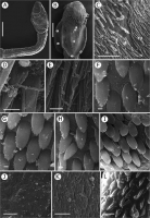
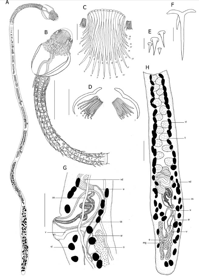 Fig. 1. Line drawings of Coronocestus ehsanentezarii sp. nov. from Iago omanensis. A Complete specimen; B Scolex, dorso-ventral view; C Apical hooks, dorso-ventral view; D Dorsal and ventral groups of lateral hooklets; E Spines on the corona; F Spine on the cephalic peduncle; G, Detail of terminal genitalia, lateral view; H, Mature proglottid, frontal view. Scale-bars: A, B: 300 μm; C: 30 μm; D, F: 20 μm; E: 10 μm; G, H: 100 μm. Abbreviations: cs cirrus sac; mg Mehlis gland; o ovary; os osmoregulatory canal;
sr seminal receptacle; t testis; u uterus; v vagina; vd vas deferens; vi vitelline follicle
Fig. 1. Line drawings of Coronocestus ehsanentezarii sp. nov. from Iago omanensis. A Complete specimen; B Scolex, dorso-ventral view; C Apical hooks, dorso-ventral view; D Dorsal and ventral groups of lateral hooklets; E Spines on the corona; F Spine on the cephalic peduncle; G, Detail of terminal genitalia, lateral view; H, Mature proglottid, frontal view. Scale-bars: A, B: 300 μm; C: 30 μm; D, F: 20 μm; E: 10 μm; G, H: 100 μm. Abbreviations: cs cirrus sac; mg Mehlis gland; o ovary; os osmoregulatory canal;
sr seminal receptacle; t testis; u uterus; v vagina; vd vas deferens; vi vitelline follicle  Fig. 2. Scanning electron micrographs of Coronocestus ehsanentezarii sp. nov. from Iago omanensis. A Scolex; B Scolex proper (note that small letters indicate locations of details shown in CI); C, D Anteriormost distal bothrial surface; E Distal bothrial surface; FH Proximal bothrial surface; I Medial line of posteriormost proximal surfaces; J Surface of cephalic peduncle; K Proglottid; L Uncinate spinitriches on cirrus. Scale-bars: A: 200 μm; B: 100 μm, C: 10 μm; DL: 1 μm
 Best viewed in Firefox


