Line Drawing 1

Fig. 1 Bothriocephalus timii n. sp. ex Cottoperca gobio. A, Entire worm showing segmentation and proglottisation of strobila (holotype IPCAS C-646/1); B, One mature proglottis per segment; vitelline f... MoreFig. 1 Bothriocephalus timii n. sp. ex Cottoperca gobio. A, Entire worm showing segmentation and proglottisation of strobila (holotype IPCAS C-646/1); B, One mature proglottis per segment; vitelline follicles only illustrated in anterior proglottis (paratype IPCAS C-646/2); C, Two mature proglottides per segment; vitelline follicles only illustrated in anterior proglottis (paratype IPCAS C-646/4). Abbreviations: an, anlagen; ad, apical disk; bo, posteriorly opened-bothria; cs, cirrus-sac; is, immagure segment; ov, ovary; t, testes; up, uterine pore; ut, uterus; vc, vaginal canal; vf, vitelline follicles. |
Line Drawing 2

Fig. 2 Bothriocephalus timii n. sp. ex Cottoperca gobio. A, Transverse section of mature proglottis at level of cirrus-sac and uterus (paratype IPCAS C-646/4); B, Transverse section of mature proglott... MoreFig. 2 Bothriocephalus timii n. sp. ex Cottoperca gobio. A, Transverse section of mature proglottis at level of cirrus-sac and uterus (paratype IPCAS C-646/4); B, Transverse section of mature proglottis at level of cirrus-sac and uterus (paratype MACN-Pa 569/1A-C); C, Transverse section of mature proglottis at level of ovary (paratype MACN-Pa 569/1A-C); D, Sagittal section of a piece of gravid proglottide (paratype IPCAS C-646/3); E, Piece of immature strobila showing osmoregulatory canals (paratype IPCAS C-646/4); F, Intrauterine egg. Abbreviations: cs, cirrus-sac; gp, genital pore; lm, bundles of longitudinal musculature; oc, osmoregulatory canals; op, operculum; ov, ovary; t, testes; up, uterine pore; ut, uterus; vc, vaginal canal; vf, vitelline follicles; vr, vitelline reservoir. |
Photo Micrograph
|
Scanning Electron Micrograph
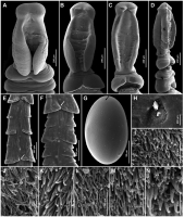
Fig. 3 Bothriocephalus timii n. sp. ex Cottoperca gobio, scanning electron micrographs. A-C, dorsoventral view of scoleces showing bothria opened posteriorly andn different state of contraction of lat... MoreFig. 3 Bothriocephalus timii n. sp. ex Cottoperca gobio, scanning electron micrographs. A-C, dorsoventral view of scoleces showing bothria opened posteriorly andn different state of contraction of lateral lappets; D, Lateral view of scolex, E, Piece of mature strobila showing external segmentation and posteriolateral wing-like expansions, with medial notches on dorsal and ventral surfaces; F, Piece of gravid strobila; G, Intrauterine egg showing operculum; H, Genital pore showing genital atrium; I-N, Surfaces shown in D at high magnification: I, Apical disk surface; J, Luminal surface of bothria; K, Lateral surface of scolex; L, Posterior surface of scolex projecting over first segment; M, Anterior surface of immature proglottis; N, Posterior surface of immature proglottis. White arrows indicate the notches of segments; black arrow indicates egg operculum. |
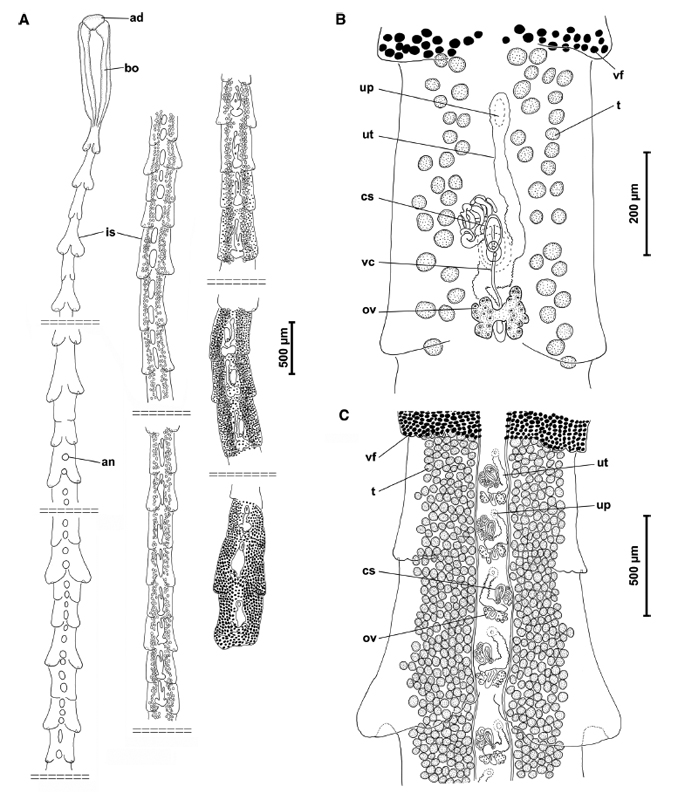 Fig. 1 Bothriocephalus timii n. sp. ex Cottoperca gobio. A, Entire worm showing segmentation and proglottisation of strobila (holotype IPCAS C-646/1); B, One mature proglottis per segment; vitelline follicles only illustrated in anterior proglottis (paratype IPCAS C-646/2); C, Two mature proglottides per segment; vitelline follicles only illustrated in anterior proglottis (paratype IPCAS C-646/4). Abbreviations: an, anlagen; ad, apical disk; bo, posteriorly opened-bothria; cs, cirrus-sac; is, immagure segment; ov, ovary; t, testes; up, uterine pore; ut, uterus; vc, vaginal canal; vf, vitelline follicles.
 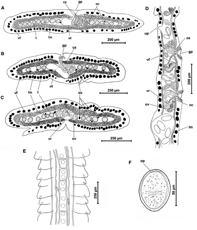 Fig. 2 Bothriocephalus timii n. sp. ex Cottoperca gobio. A, Transverse section of mature proglottis at level of cirrus-sac and uterus (paratype IPCAS C-646/4); B, Transverse section of mature proglottis at level of cirrus-sac and uterus (paratype MACN-Pa 569/1A-C); C, Transverse section of mature proglottis at level of ovary (paratype MACN-Pa 569/1A-C); D, Sagittal section of a piece of gravid proglottide (paratype IPCAS C-646/3); E, Piece of immature strobila showing osmoregulatory canals (paratype IPCAS C-646/4); F, Intrauterine egg. Abbreviations: cs, cirrus-sac; gp, genital pore; lm, bundles of longitudinal musculature; oc, osmoregulatory canals; op, operculum; ov, ovary; t, testes; up, uterine pore; ut, uterus; vc, vaginal canal; vf, vitelline follicles; vr, vitelline reservoir.
  Fig. 3 Bothriocephalus timii n. sp. ex Cottoperca gobio, scanning electron micrographs. A-C, dorsoventral view of scoleces showing bothria opened posteriorly andn different state of contraction of lateral lappets; D, Lateral view of scolex, E, Piece of mature strobila showing external segmentation and posteriolateral wing-like expansions, with medial notches on dorsal and ventral surfaces; F, Piece of gravid strobila; G, Intrauterine egg showing operculum; H, Genital pore showing genital atrium; I-N, Surfaces shown in D at high magnification: I, Apical disk surface; J, Luminal surface of bothria; K, Lateral surface of scolex; L, Posterior surface of scolex projecting over first segment; M, Anterior surface of immature proglottis; N, Posterior surface of immature proglottis. White arrows indicate the notches of segments; black arrow indicates egg operculum.

