Line Drawing 1

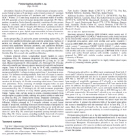
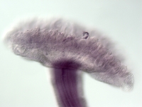
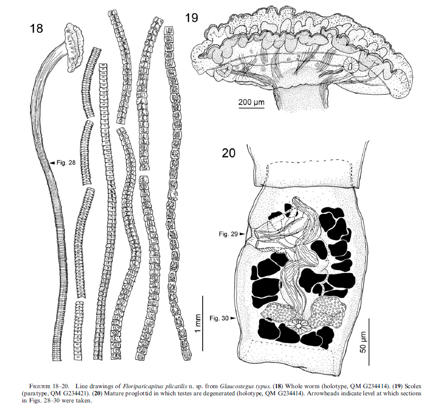
FIGURES 1820. Line drawings of Floriparicapitus plicatilis n. sp. from Glaucostegus typus. (18) Whole worm (holotype, QM G234414). (19) Scolex
(paratype, QM G234421). (20) Mature proglottid in which... MoreFIGURES 1820. Line drawings of Floriparicapitus plicatilis n. sp. from Glaucostegus typus. (18) Whole worm (holotype, QM G234414). (19) Scolex
(paratype, QM G234421). (20) Mature proglottid in which testes are degenerated (holotype, QM G234414). Arrowheads indicate level at which sections in Figs. 2830 were taken. |
Line Drawing 2
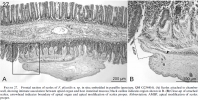
FIGURE 27. Frontal section of scolex of F. plicatilis n. sp. in situ; embedded in paraffin (paratype, QM G234416). (A) Scolex attached to chamber
wall, showing intimate association between apical org... MoreFIGURE 27. Frontal section of scolex of F. plicatilis n. sp. in situ; embedded in paraffin (paratype, QM G234416). (A) Scolex attached to chamber
wall, showing intimate association between apical organ and host intestinal mucosa; black outline indicates region shown in B. (B) Close-up of attached
scolex; arrowhead indicates boundary of apical organ and apical modification of scolex proper. Abbreviation: AMSP, apical modification of scolex
proper. |
Photo Micrograph
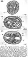
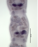
FIGURES 2830. Proglottid cross-sections of F. plicatilis n. sp.;
embedded in paraffin. (28) Through anterior region of strobila showing
six excretory vessels (arrowheads) and discrete longitudinal ... MoreFIGURES 2830. Proglottid cross-sections of F. plicatilis n. sp.;
embedded in paraffin. (28) Through anterior region of strobila showing
six excretory vessels (arrowheads) and discrete longitudinal muscle
bundles (paratype, QM G234416). (29) Through mature proglottid at
level of genital pore. Note vagina and cirrus opening at approximately
same level (paratype, USNPC 108159). (30) Through mature proglottid at
level of ovarian bridge (paratype, USNPC 108159). Note expanded size of
medial pair of excretory vessels (arrowheads). Abbreviations: C, cirrus;
CS, cirrus sac; GP, genital pore; LM, longitudinal muscle bundles; MG,
Mehlis gland; OV, ovary; OC, ovicapt; OD, oviduct; T, testis; U, uterus;
UD, uterine duct; VA, vagina; VF, vitelline follicle; VD, vas deferens. |
Scanning Electron Micrograph
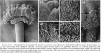
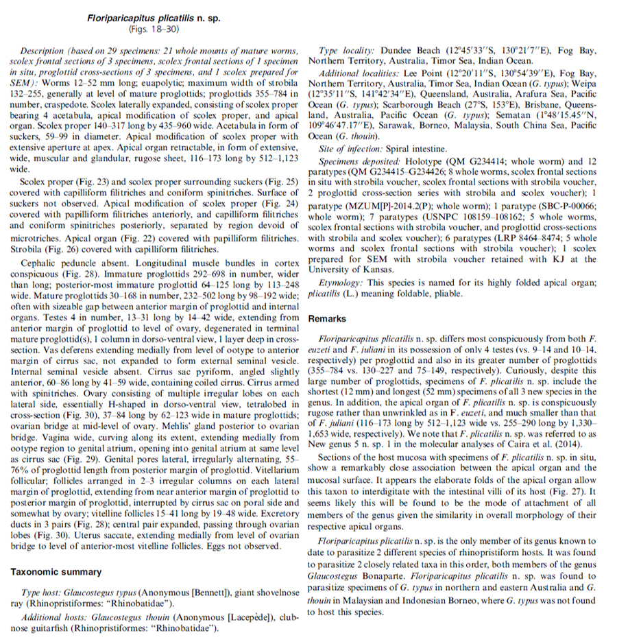
FIGURES 2126. Scanning electron micrographs of F. plicatilis n. sp. from G. typus. (21) Scolex; small numbers indicate location of detail in Figs. 22
26. (22) Apical organ with papilliform filitric... MoreFIGURES 2126. Scanning electron micrographs of F. plicatilis n. sp. from G. typus. (21) Scolex; small numbers indicate location of detail in Figs. 22
26. (22) Apical organ with papilliform filitriches. (23) Scolex proper between suckers with capilliform filitriches and coniform spinitriches. (24)
Microthrix variation of apical modification of scolex proper (AMSP); anterior AMSP with papilliform filitriches and posterior AMSP with capilliform
filitriches and coniform spinitriches, separated by region devoid of microtriches. (25) Scolex proper surrounding suckers with capilliform filitriches and coniform spinitriches. (26) Strobila with capilliform filitriches. |

 FIGURES 1820. Line drawings of Floriparicapitus plicatilis n. sp. from Glaucostegus typus. (18) Whole worm (holotype, QM G234414). (19) Scolex
(paratype, QM G234421). (20) Mature proglottid in which testes are degenerated (holotype, QM G234414). Arrowheads indicate level at which sections in Figs. 2830 were taken.
FIGURES 1820. Line drawings of Floriparicapitus plicatilis n. sp. from Glaucostegus typus. (18) Whole worm (holotype, QM G234414). (19) Scolex
(paratype, QM G234421). (20) Mature proglottid in which testes are degenerated (holotype, QM G234414). Arrowheads indicate level at which sections in Figs. 2830 were taken. 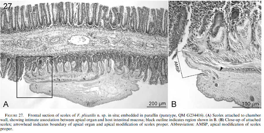 FIGURE 27. Frontal section of scolex of F. plicatilis n. sp. in situ; embedded in paraffin (paratype, QM G234416). (A) Scolex attached to chamber
wall, showing intimate association between apical organ and host intestinal mucosa; black outline indicates region shown in B. (B) Close-up of attached
scolex; arrowhead indicates boundary of apical organ and apical modification of scolex proper. Abbreviation: AMSP, apical modification of scolex
proper.
FIGURE 27. Frontal section of scolex of F. plicatilis n. sp. in situ; embedded in paraffin (paratype, QM G234416). (A) Scolex attached to chamber
wall, showing intimate association between apical organ and host intestinal mucosa; black outline indicates region shown in B. (B) Close-up of attached
scolex; arrowhead indicates boundary of apical organ and apical modification of scolex proper. Abbreviation: AMSP, apical modification of scolex
proper.  FIGURES 2830. Proglottid cross-sections of F. plicatilis n. sp.;
embedded in paraffin. (28) Through anterior region of strobila showing
six excretory vessels (arrowheads) and discrete longitudinal muscle
bundles (paratype, QM G234416). (29) Through mature proglottid at
level of genital pore. Note vagina and cirrus opening at approximately
same level (paratype, USNPC 108159). (30) Through mature proglottid at
level of ovarian bridge (paratype, USNPC 108159). Note expanded size of
medial pair of excretory vessels (arrowheads). Abbreviations: C, cirrus;
CS, cirrus sac; GP, genital pore; LM, longitudinal muscle bundles; MG,
Mehlis gland; OV, ovary; OC, ovicapt; OD, oviduct; T, testis; U, uterus;
UD, uterine duct; VA, vagina; VF, vitelline follicle; VD, vas deferens.
FIGURES 2830. Proglottid cross-sections of F. plicatilis n. sp.;
embedded in paraffin. (28) Through anterior region of strobila showing
six excretory vessels (arrowheads) and discrete longitudinal muscle
bundles (paratype, QM G234416). (29) Through mature proglottid at
level of genital pore. Note vagina and cirrus opening at approximately
same level (paratype, USNPC 108159). (30) Through mature proglottid at
level of ovarian bridge (paratype, USNPC 108159). Note expanded size of
medial pair of excretory vessels (arrowheads). Abbreviations: C, cirrus;
CS, cirrus sac; GP, genital pore; LM, longitudinal muscle bundles; MG,
Mehlis gland; OV, ovary; OC, ovicapt; OD, oviduct; T, testis; U, uterus;
UD, uterine duct; VA, vagina; VF, vitelline follicle; VD, vas deferens. 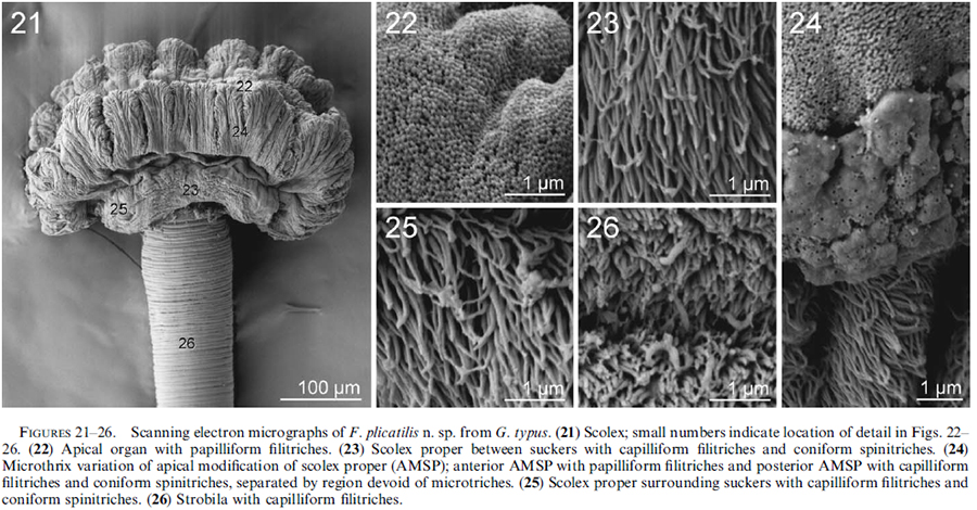 FIGURES 2126. Scanning electron micrographs of F. plicatilis n. sp. from G. typus. (21) Scolex; small numbers indicate location of detail in Figs. 22
26. (22) Apical organ with papilliform filitriches. (23) Scolex proper between suckers with capilliform filitriches and coniform spinitriches. (24)
Microthrix variation of apical modification of scolex proper (AMSP); anterior AMSP with papilliform filitriches and posterior AMSP with capilliform
filitriches and coniform spinitriches, separated by region devoid of microtriches. (25) Scolex proper surrounding suckers with capilliform filitriches and coniform spinitriches. (26) Strobila with capilliform filitriches.
FIGURES 2126. Scanning electron micrographs of F. plicatilis n. sp. from G. typus. (21) Scolex; small numbers indicate location of detail in Figs. 22
26. (22) Apical organ with papilliform filitriches. (23) Scolex proper between suckers with capilliform filitriches and coniform spinitriches. (24)
Microthrix variation of apical modification of scolex proper (AMSP); anterior AMSP with papilliform filitriches and posterior AMSP with capilliform
filitriches and coniform spinitriches, separated by region devoid of microtriches. (25) Scolex proper surrounding suckers with capilliform filitriches and coniform spinitriches. (26) Strobila with capilliform filitriches. 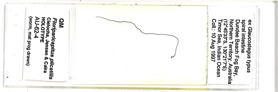 Floriparicapitus plicatilis holotype
QMG234414
(AU-62-4)
Floriparicapitus plicatilis holotype
QMG234414
(AU-62-4)  Floriparicapitus plicatilis holotype
QMG234414
(AU-62-4)
Floriparicapitus plicatilis holotype
QMG234414
(AU-62-4)  Floriparicapitus plicatilis paratype
G234421
(AU-62-3)
Floriparicapitus plicatilis paratype
G234421
(AU-62-3) 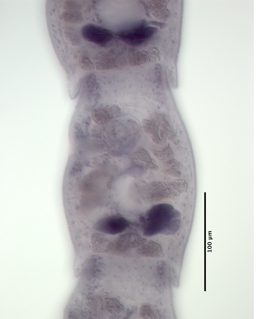 Floriparicapitus plicatilis paratype
G234421
(AU-62-3)
Floriparicapitus plicatilis paratype
G234421
(AU-62-3) 

