Line Drawing 1
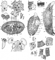
PLATE 1. FIGURES 1-6. Phyllbothrium tumidum n. sp. 1. Scolex; dorso-ventral view; drawn from an alcoholic specimen. Actual diameter 2.1 mm. 2. Front view of scolex mounted in balsam; partly diagra... MorePLATE 1. FIGURES 1-6. Phyllbothrium tumidum n. sp. 1. Scolex; dorso-ventral view; drawn from an alcoholic specimen. Actual diameter 2.1 mm. 2. Front view of scolex mounted in balsam; partly diagrammatic, folds which partly covered the auxiliary suckers, having been omitted; greatest diameter 4.6 mm. 3. Single bothrium of scolex fixed while attached to mucous membrane of host, mounted in balsam; diameter of auxiliary sucker 0.35 mm. 4. Proglottides, 15 mm. from scolex; balsam; breadth 1.5 mm. 5. Posterior ends of three stobiles; balsam; length of longest 9 mm. 6. Transverse section of mature, but unripe, proglottis, a little in front of the genital aperture; greatest diameter 1 mm.
PLATE 2. FIGURES 7-10. Phyllbothrium tumidum n. sp. 7. Free proglottis, mature but unripe; balsam; length 5 mm. Sketch by George T. Kline. 8. Free, ripe proglottis; balsam; lenth 7.5 mm. 9. Diagram showing relation of ducts of reproductive organs in vicintiy of the shell gland, new species. 10. Detail of longitudinal muscles in cross section, figure 6.
PLATE 3. FIGURES 11-15. Phyllbothrium tumidum n. sp. 11. Sagittal section, somewhat slanting, of anterior end of scolex, showing ganglion cells and excretory vessels; diameter of auxiliary sucker 0.25 mm. 12. Ganglion cells in sections of scolex. a, cells in parenchyma; b, black of bothrium, and cells associated with muscle fibers; d, cells associated with muscle fibers at base of pedicel; e, marginal region of bothrium; f, central region of bothrium; camera lucida drawings with Spencer NO.6 ocular, and o.4 mm. objective. 13. Sagittal section of cuticle. See text. 14. Larval Phyllobthrium, from swordfish; balsam; length 1.54 mm. 15. Larval Phyllbothrium from swordfish; much flattened at time of fixing; balsam; breadth of neck at base of scolex 0.75 mm. Abbreviations: ci, cirrus; cp, cirrus-pouch; ed, dorsal excretory vessel; ev, ventral excretory vessel; ga, genital aperature; gd, germ duct; lm, longitudinal muscle; n, nerve; o, ovary (germarium); sd, sperm duct; sg, shell gland; t, testes; u,uterus; v, vagina; vd, vas deferens; vg, vitelline gland; yd, vitelline duct. |
Line Drawing 2
|
Photo Micrograph
|
Scanning Electron Micrograph
|
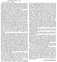
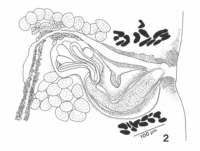
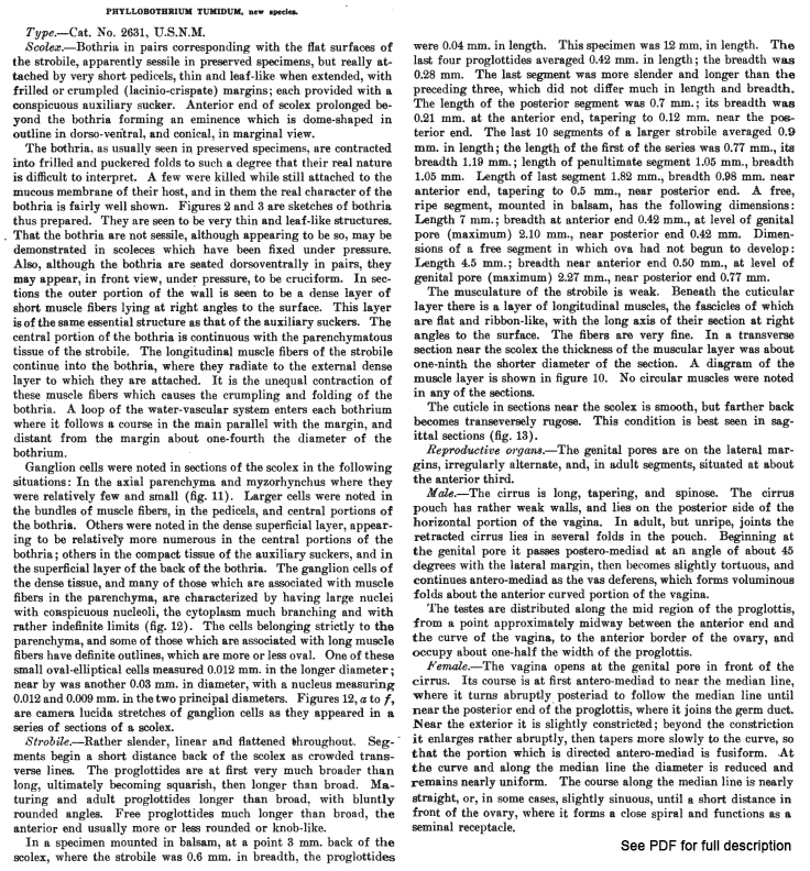
 PLATE 1. FIGURES 1-6. Phyllbothrium tumidum n. sp. 1. Scolex; dorso-ventral view; drawn from an alcoholic specimen. Actual diameter 2.1 mm. 2. Front view of scolex mounted in balsam; partly diagrammatic, folds which partly covered the auxiliary suckers, having been omitted; greatest diameter 4.6 mm. 3. Single bothrium of scolex fixed while attached to mucous membrane of host, mounted in balsam; diameter of auxiliary sucker 0.35 mm. 4. Proglottides, 15 mm. from scolex; balsam; breadth 1.5 mm. 5. Posterior ends of three stobiles; balsam; length of longest 9 mm. 6. Transverse section of mature, but unripe, proglottis, a little in front of the genital aperture; greatest diameter 1 mm.
PLATE 2. FIGURES 7-10. Phyllbothrium tumidum n. sp. 7. Free proglottis, mature but unripe; balsam; length 5 mm. Sketch by George T. Kline. 8. Free, ripe proglottis; balsam; lenth 7.5 mm. 9. Diagram showing relation of ducts of reproductive organs in vicintiy of the shell gland, new species. 10. Detail of longitudinal muscles in cross section, figure 6.
PLATE 3. FIGURES 11-15. Phyllbothrium tumidum n. sp. 11. Sagittal section, somewhat slanting, of anterior end of scolex, showing ganglion cells and excretory vessels; diameter of auxiliary sucker 0.25 mm. 12. Ganglion cells in sections of scolex. a, cells in parenchyma; b, black of bothrium, and cells associated with muscle fibers; d, cells associated with muscle fibers at base of pedicel; e, marginal region of bothrium; f, central region of bothrium; camera lucida drawings with Spencer NO.6 ocular, and o.4 mm. objective. 13. Sagittal section of cuticle. See text. 14. Larval Phyllobthrium, from swordfish; balsam; length 1.54 mm. 15. Larval Phyllbothrium from swordfish; much flattened at time of fixing; balsam; breadth of neck at base of scolex 0.75 mm. Abbreviations: ci, cirrus; cp, cirrus-pouch; ed, dorsal excretory vessel; ev, ventral excretory vessel; ga, genital aperature; gd, germ duct; lm, longitudinal muscle; n, nerve; o, ovary (germarium); sd, sperm duct; sg, shell gland; t, testes; u,uterus; v, vagina; vd, vas deferens; vg, vitelline gland; yd, vitelline duct.
PLATE 1. FIGURES 1-6. Phyllbothrium tumidum n. sp. 1. Scolex; dorso-ventral view; drawn from an alcoholic specimen. Actual diameter 2.1 mm. 2. Front view of scolex mounted in balsam; partly diagrammatic, folds which partly covered the auxiliary suckers, having been omitted; greatest diameter 4.6 mm. 3. Single bothrium of scolex fixed while attached to mucous membrane of host, mounted in balsam; diameter of auxiliary sucker 0.35 mm. 4. Proglottides, 15 mm. from scolex; balsam; breadth 1.5 mm. 5. Posterior ends of three stobiles; balsam; length of longest 9 mm. 6. Transverse section of mature, but unripe, proglottis, a little in front of the genital aperture; greatest diameter 1 mm.
PLATE 2. FIGURES 7-10. Phyllbothrium tumidum n. sp. 7. Free proglottis, mature but unripe; balsam; length 5 mm. Sketch by George T. Kline. 8. Free, ripe proglottis; balsam; lenth 7.5 mm. 9. Diagram showing relation of ducts of reproductive organs in vicintiy of the shell gland, new species. 10. Detail of longitudinal muscles in cross section, figure 6.
PLATE 3. FIGURES 11-15. Phyllbothrium tumidum n. sp. 11. Sagittal section, somewhat slanting, of anterior end of scolex, showing ganglion cells and excretory vessels; diameter of auxiliary sucker 0.25 mm. 12. Ganglion cells in sections of scolex. a, cells in parenchyma; b, black of bothrium, and cells associated with muscle fibers; d, cells associated with muscle fibers at base of pedicel; e, marginal region of bothrium; f, central region of bothrium; camera lucida drawings with Spencer NO.6 ocular, and o.4 mm. objective. 13. Sagittal section of cuticle. See text. 14. Larval Phyllobthrium, from swordfish; balsam; length 1.54 mm. 15. Larval Phyllbothrium from swordfish; much flattened at time of fixing; balsam; breadth of neck at base of scolex 0.75 mm. Abbreviations: ci, cirrus; cp, cirrus-pouch; ed, dorsal excretory vessel; ev, ventral excretory vessel; ga, genital aperature; gd, germ duct; lm, longitudinal muscle; n, nerve; o, ovary (germarium); sd, sperm duct; sg, shell gland; t, testes; u,uterus; v, vagina; vd, vas deferens; vg, vitelline gland; yd, vitelline duct.  From Ruhnke, 1993 (Cit# 389). Fig. 2. Cirrus sac from end segment of lectotype (USNM no. 7631) of C. tumidum n. comb.


