Line Drawing 1
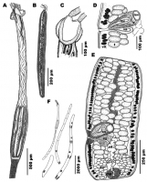
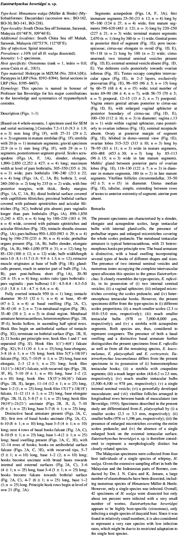
Line drawings of Eutetrarhynchus beveridgei n. sp. from Himantura walga (Mu¨ller & Henle) (BO-141: B, C, E; BO-162: A, F; BO-238: D) in the South China Sea. A, scolex, lateral view; B, bulb; C, pars b... MoreLine drawings of Eutetrarhynchus beveridgei n. sp. from Himantura walga (Mu¨ller & Henle) (BO-141: B, C, E; BO-162: A, F; BO-238: D) in the South China Sea. A, scolex, lateral view; B, bulb; C, pars bothrialis, dorso-ventral view; D, detail of cirrus-sac, composite line drawing from serial sections; E, mature segment; F, outline of entire cestode. Abbreviations: c, cirrus; cs, cirrus-sac; isv, internal seminal vesicle; mg, Mehlis gland; ov, ovary; t, testis; va, vagina; vit, vitelline follicle; vs, vaginal sphincter |
Line Drawing 2
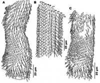
Line drawings of tentacular armature of Eutetrarhynchus beveridgei n. sp. from Himantura walga (Mu¨ller& Henle) (BO-30: C; BO-162: A, B) in the South China Sea. A, basal tentacular armature, internal ... MoreLine drawings of tentacular armature of Eutetrarhynchus beveridgei n. sp. from Himantura walga (Mu¨ller& Henle) (BO-30: C; BO-162: A, B) in the South China Sea. A, basal tentacular armature, internal surface; B, metabasal tentacular armature, bothrial to external surface; C, basal tentacular armature, antibothrial to external surface |
Photo Micrograph
|
Scanning Electron Micrograph
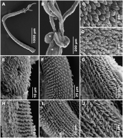
Scanning electron micrographs of Eutetrarhynchus beveridgei n. sp. from Himantura walga (Mu¨ller & Henle) (BO-238) in the South China Sea. A, complete specimen, lateral view; B, detailed view of pars ... MoreScanning electron micrographs of Eutetrarhynchus beveridgei n. sp. from Himantura walga (Mu¨ller & Henle) (BO-238) in the South China Sea. A, complete specimen, lateral view; B, detailed view of pars bothrialis; C, palmate spinitriches and acicular filitriches covering proximal bothrial surface; D, palmate spinitriches and acicular filitriches covering scolex peduncle; E, basal tentacular armature, bothrial surface; F, metabasal tentacular armature, external surface; G, metabasal tentacular armature, antibothrial surface; H, metabasal tentacular armature, internal surface; I, metabasal tentacular armature, bothrial surface; J, metabasal tentacular armature, close-up on bothrial surface. Symbols: *, position on proximal bothrial surface where microthrix micrograph (C) was taken; +, position position on scolex peduncle where microthrix micrograph (D) was taken on scolex peduncle where microthrix micrograph |
 Line drawings of Eutetrarhynchus beveridgei n. sp. from Himantura walga (Mu¨ller & Henle) (BO-141: B, C, E; BO-162: A, F; BO-238: D) in the South China Sea. A, scolex, lateral view; B, bulb; C, pars bothrialis, dorso-ventral view; D, detail of cirrus-sac, composite line drawing from serial sections; E, mature segment; F, outline of entire cestode. Abbreviations: c, cirrus; cs, cirrus-sac; isv, internal seminal vesicle; mg, Mehlis gland; ov, ovary; t, testis; va, vagina; vit, vitelline follicle; vs, vaginal sphincter
 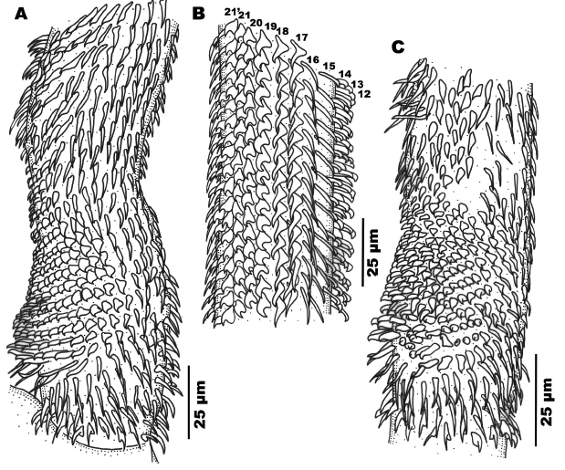 Line drawings of tentacular armature of Eutetrarhynchus beveridgei n. sp. from Himantura walga (Mu¨ller& Henle) (BO-30: C; BO-162: A, B) in the South China Sea. A, basal tentacular armature, internal surface; B, metabasal tentacular armature, bothrial to external surface; C, basal tentacular armature, antibothrial to external surface
 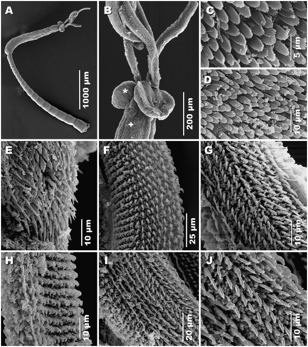 Scanning electron micrographs of Eutetrarhynchus beveridgei n. sp. from Himantura walga (Mu¨ller & Henle) (BO-238) in the South China Sea. A, complete specimen, lateral view; B, detailed view of pars bothrialis; C, palmate spinitriches and acicular filitriches covering proximal bothrial surface; D, palmate spinitriches and acicular filitriches covering scolex peduncle; E, basal tentacular armature, bothrial surface; F, metabasal tentacular armature, external surface; G, metabasal tentacular armature, antibothrial surface; H, metabasal tentacular armature, internal surface; I, metabasal tentacular armature, bothrial surface; J, metabasal tentacular armature, close-up on bothrial surface. Symbols: *, position on proximal bothrial surface where microthrix micrograph (C) was taken; +, position position on scolex peduncle where microthrix micrograph (D) was taken on scolex peduncle where microthrix micrograph

