Line Drawing 1

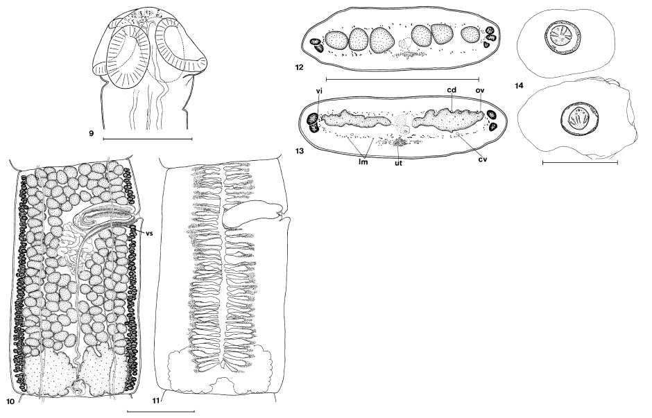
Figures 911. Nomimoscolex pertierrae n. sp. 9. Paratype BMNH 2005.10.3.4, scolex. 10. Holotype IOC 36598, pregravid proglottis,
ventral view (see Figure 11 for uterus). 11. Holotype IOC 36598, same ... MoreFigures 911. Nomimoscolex pertierrae n. sp. 9. Paratype BMNH 2005.10.3.4, scolex. 10. Holotype IOC 36598, pregravid proglottis,
ventral view (see Figure 11 for uterus). 11. Holotype IOC 36598, same proglottis as figure 10, but only the uterus is figured.
Abbreviation: vs, vaginal sphincter. Scale-bars: 9, 250 µm; 10,11, 500 µm. Figures 1214. Nomimoscolex pertierrae n. sp. 12, 13. Paratype INVE 37174, cross-section of a premature proglottis. 12. Crosssection
at the posterior level of a proglottis. 13. Cross-section at the level of the ovary. 14. Eggs drawn in distilled water. Abbreviations:
cd, dorsal osmoregulatory canal; cv, ventral osmoregulatory canal; lm, internal longitudinal musculature; ov, ovary; ut,
uterus; vi, vitelline follicles. Scale-bars: 12,13, 500 µm; 14, 50 µm. |
Line Drawing 2
|
Photo Micrograph
|
Scanning Electron Micrograph
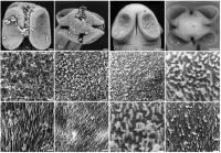
Figures 1520. Nomimoscolex sudobim Woodland, 1935. Scanning electron micrographs of the scolex, INVE 19448. 15. Dorsoventral
view. 16. Apical view. 17. Detail of the apex showing the compact filifor... MoreFigures 1520. Nomimoscolex sudobim Woodland, 1935. Scanning electron micrographs of the scolex, INVE 19448. 15. Dorsoventral
view. 16. Apical view. 17. Detail of the apex showing the compact filiform microtriches. 18. Upper margin of sucker, with
filiform microtriches. 19. Internal surface of the sucker, showing ensiform microtriches. 20. Proliferation zone, ensiform microtriches.
Scale-bars: 15,16, 50 µm; 1720, 2 µm.
Figures 2126. Nomimoscolex pertierrae n. sp. Scanning electron micrographs of the scolex, INVE 37175. 21. Dorso-ventral view.
22. Apical view. 23. Detail of the apex showing the papilliform to short filiform microtriches. 24. Upper margin of the sucker, with
slender digitiform microtriches. 25. Internal surface of the sucker, with slender digitiform microtriches. 26. Proliferation zone, with
ensiform microtriches. Scale-bars: 21,22, 50 µm; 23,26, 2 µm; 24,25, 1 µm. |
 Figures 911. Nomimoscolex pertierrae n. sp. 9. Paratype BMNH 2005.10.3.4, scolex. 10. Holotype IOC 36598, pregravid proglottis,
ventral view (see Figure 11 for uterus). 11. Holotype IOC 36598, same proglottis as figure 10, but only the uterus is figured.
Abbreviation: vs, vaginal sphincter. Scale-bars: 9, 250 µm; 10,11, 500 µm. Figures 1214. Nomimoscolex pertierrae n. sp. 12, 13. Paratype INVE 37174, cross-section of a premature proglottis. 12. Crosssection
at the posterior level of a proglottis. 13. Cross-section at the level of the ovary. 14. Eggs drawn in distilled water. Abbreviations:
cd, dorsal osmoregulatory canal; cv, ventral osmoregulatory canal; lm, internal longitudinal musculature; ov, ovary; ut,
uterus; vi, vitelline follicles. Scale-bars: 12,13, 500 µm; 14, 50 µm.
Figures 911. Nomimoscolex pertierrae n. sp. 9. Paratype BMNH 2005.10.3.4, scolex. 10. Holotype IOC 36598, pregravid proglottis,
ventral view (see Figure 11 for uterus). 11. Holotype IOC 36598, same proglottis as figure 10, but only the uterus is figured.
Abbreviation: vs, vaginal sphincter. Scale-bars: 9, 250 µm; 10,11, 500 µm. Figures 1214. Nomimoscolex pertierrae n. sp. 12, 13. Paratype INVE 37174, cross-section of a premature proglottis. 12. Crosssection
at the posterior level of a proglottis. 13. Cross-section at the level of the ovary. 14. Eggs drawn in distilled water. Abbreviations:
cd, dorsal osmoregulatory canal; cv, ventral osmoregulatory canal; lm, internal longitudinal musculature; ov, ovary; ut,
uterus; vi, vitelline follicles. Scale-bars: 12,13, 500 µm; 14, 50 µm. 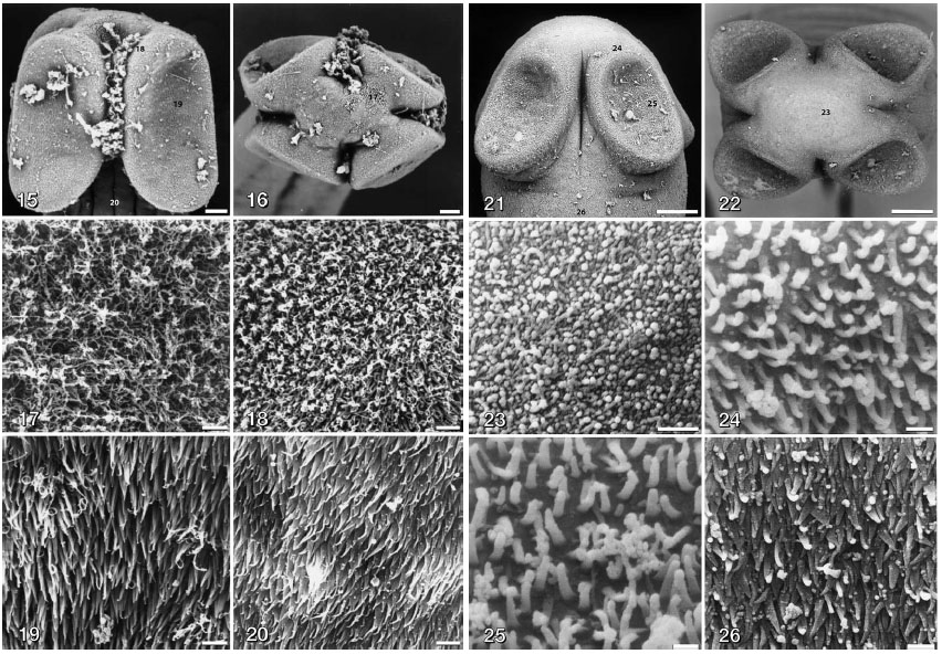 Figures 1520. Nomimoscolex sudobim Woodland, 1935. Scanning electron micrographs of the scolex, INVE 19448. 15. Dorsoventral
view. 16. Apical view. 17. Detail of the apex showing the compact filiform microtriches. 18. Upper margin of sucker, with
filiform microtriches. 19. Internal surface of the sucker, showing ensiform microtriches. 20. Proliferation zone, ensiform microtriches.
Scale-bars: 15,16, 50 µm; 1720, 2 µm.
Figures 2126. Nomimoscolex pertierrae n. sp. Scanning electron micrographs of the scolex, INVE 37175. 21. Dorso-ventral view.
22. Apical view. 23. Detail of the apex showing the papilliform to short filiform microtriches. 24. Upper margin of the sucker, with
slender digitiform microtriches. 25. Internal surface of the sucker, with slender digitiform microtriches. 26. Proliferation zone, with
ensiform microtriches. Scale-bars: 21,22, 50 µm; 23,26, 2 µm; 24,25, 1 µm.
Figures 1520. Nomimoscolex sudobim Woodland, 1935. Scanning electron micrographs of the scolex, INVE 19448. 15. Dorsoventral
view. 16. Apical view. 17. Detail of the apex showing the compact filiform microtriches. 18. Upper margin of sucker, with
filiform microtriches. 19. Internal surface of the sucker, showing ensiform microtriches. 20. Proliferation zone, ensiform microtriches.
Scale-bars: 15,16, 50 µm; 1720, 2 µm.
Figures 2126. Nomimoscolex pertierrae n. sp. Scanning electron micrographs of the scolex, INVE 37175. 21. Dorso-ventral view.
22. Apical view. 23. Detail of the apex showing the papilliform to short filiform microtriches. 24. Upper margin of the sucker, with
slender digitiform microtriches. 25. Internal surface of the sucker, with slender digitiform microtriches. 26. Proliferation zone, with
ensiform microtriches. Scale-bars: 21,22, 50 µm; 23,26, 2 µm; 24,25, 1 µm. 

