Line Drawing 1
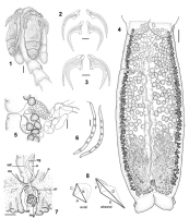
FIGURES 18. Calliobothrium australis. 1. Scolex. Bar 50 µm. 2. Detail of hooks. Bar 25 µm. 3. Detail of hooks. Bar 25 µm. 4. Mature segment. Bar 200 µm. 5. Detail of terminal genitalia. Bar 40 µm. 6.... MoreFIGURES 18. Calliobothrium australis. 1. Scolex. Bar 50 µm. 2. Detail of hooks. Bar 25 µm. 3. Detail of hooks. Bar 25 µm. 4. Mature segment. Bar 200 µm. 5. Detail of terminal genitalia. Bar 40 µm. 6. Detail of cocoons. Bar 100 µm. 7. Detail of ootype region. Bar 100 µm. 8. Hook measurements. Abbreviations: mg, Mehlis gland; o, ovary; oc, oocapt; od, oviduct; sr, seminal receptacle; u, uterus; ud,
uteroduct; vg, vagina. |
Line Drawing 2
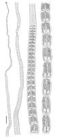
FIGURE 9. Calliobothrium australis. Entire worm. Bar 1 mm. |
Photo Micrograph

FIGURES 1921. Calliobothrium australis, cross sections of mature
segment. 19. Cross section at the level of testes anterior to cirrus sac.
20. Cross section at the level of cirrus sac. 21. Cross se... MoreFIGURES 1921. Calliobothrium australis, cross sections of mature
segment. 19. Cross section at the level of testes anterior to cirrus sac.
20. Cross section at the level of cirrus sac. 21. Cross section at the level
of ovarian isthmus. Bar 100 µm. Abbreviations: cs, cirrus sac; mg, Mehlis gland; nc, nerve cord; o, ovary; t, testis; u, uterus; vd, vas deferens; vf, vitelline follicle; vg, vagina; vod, ventral osmoregulatory duct. |
Scanning Electron Micrograph
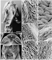
FIGURES 1018. Calliobothrium australis, scanning electron micrographs. 10. Scolex. Bar 50 µm. 11. Microtriches on proximal bothridial surface. Bar 2.5 µm. 12. Microtriches on distal bothridial surfa... MoreFIGURES 1018. Calliobothrium australis, scanning electron micrographs. 10. Scolex. Bar 50 µm. 11. Microtriches on proximal bothridial surface. Bar 2.5 µm. 12. Microtriches on distal bothridial surface. Bar 2.5 µm. 13. Detail of muscular pad showing apical suckers. Bar
10 µm. 14. Microtriches on proximal surface of muscular pad. Bar 2.5 µm. 15. Microtriches on distal surface of muscular pad. Bar 1 µm.
16. Immature segment. Bar 10 µm. 17. Detail of microtriches on anterior half of immature segment. Bar 1 µm. 18. Detail of microtriches
on flaps of immature segment. Bar 2.5 µm. |
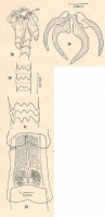
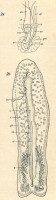
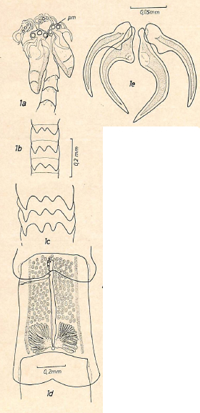 Fig. 1. Calliobothrium verticillatum australis subsp. nov. a, scolex : b, c, lacinios de los
bordes posteriores de los segmentos en distinta posicion del estrobilo; d, penultimo segmento:
e, ganchos. (be, bolsa del cirro: cd, canal deferente: cf, camara de fertillzacion ; cv,
conductos vitelogenos ; e, embrion ; gm, glandula de Mehlis ; gv, glandulas vitelogenas ; o,
ovario : og, organo glandular ; oo, ootipo : ov, oviduoto : ovc, oviscapto : pm. porcion muscular ; t, testiculos ; u, utero ; ul, uteroducto ; v, ventosa : va, vagina.)
Fig. 1. Calliobothrium verticillatum australis subsp. nov. a, scolex : b, c, lacinios de los
bordes posteriores de los segmentos en distinta posicion del estrobilo; d, penultimo segmento:
e, ganchos. (be, bolsa del cirro: cd, canal deferente: cf, camara de fertillzacion ; cv,
conductos vitelogenos ; e, embrion ; gm, glandula de Mehlis ; gv, glandulas vitelogenas ; o,
ovario : og, organo glandular ; oo, ootipo : ov, oviduoto : ovc, oviscapto : pm. porcion muscular ; t, testiculos ; u, utero ; ul, uteroducto ; v, ventosa : va, vagina.) 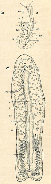 Fig. 2. Calliobothrium verticillatum australis subsp. nov. a, conductos genitales (esquematico);
b, segmento libre. (Abreviaturas como en fig. 1).
Fig. 2. Calliobothrium verticillatum australis subsp. nov. a, conductos genitales (esquematico);
b, segmento libre. (Abreviaturas como en fig. 1). 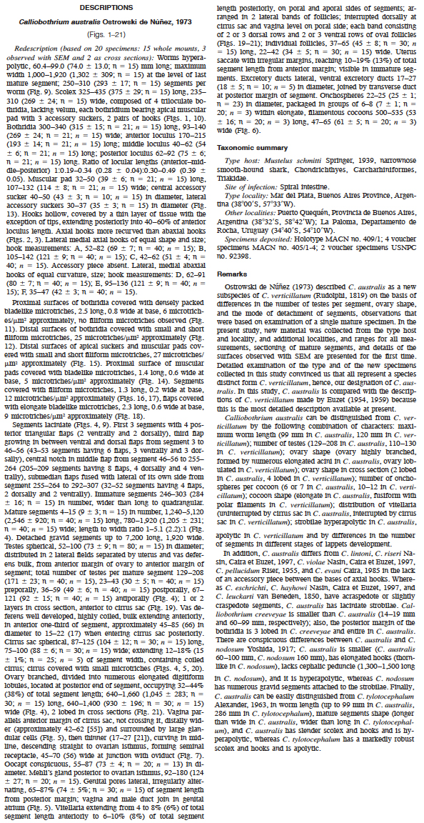
 FIGURES 18. Calliobothrium australis. 1. Scolex. Bar 50 µm. 2. Detail of hooks. Bar 25 µm. 3. Detail of hooks. Bar 25 µm. 4. Mature segment. Bar 200 µm. 5. Detail of terminal genitalia. Bar 40 µm. 6. Detail of cocoons. Bar 100 µm. 7. Detail of ootype region. Bar 100 µm. 8. Hook measurements. Abbreviations: mg, Mehlis gland; o, ovary; oc, oocapt; od, oviduct; sr, seminal receptacle; u, uterus; ud,
uteroduct; vg, vagina.
FIGURES 18. Calliobothrium australis. 1. Scolex. Bar 50 µm. 2. Detail of hooks. Bar 25 µm. 3. Detail of hooks. Bar 25 µm. 4. Mature segment. Bar 200 µm. 5. Detail of terminal genitalia. Bar 40 µm. 6. Detail of cocoons. Bar 100 µm. 7. Detail of ootype region. Bar 100 µm. 8. Hook measurements. Abbreviations: mg, Mehlis gland; o, ovary; oc, oocapt; od, oviduct; sr, seminal receptacle; u, uterus; ud,
uteroduct; vg, vagina.  FIGURE 9. Calliobothrium australis. Entire worm. Bar 1 mm.
  FIGURES 1921. Calliobothrium australis, cross sections of mature
segment. 19. Cross section at the level of testes anterior to cirrus sac.
20. Cross section at the level of cirrus sac. 21. Cross section at the level
of ovarian isthmus. Bar 100 µm. Abbreviations: cs, cirrus sac; mg, Mehlis gland; nc, nerve cord; o, ovary; t, testis; u, uterus; vd, vas deferens; vf, vitelline follicle; vg, vagina; vod, ventral osmoregulatory duct.
FIGURES 1921. Calliobothrium australis, cross sections of mature
segment. 19. Cross section at the level of testes anterior to cirrus sac.
20. Cross section at the level of cirrus sac. 21. Cross section at the level
of ovarian isthmus. Bar 100 µm. Abbreviations: cs, cirrus sac; mg, Mehlis gland; nc, nerve cord; o, ovary; t, testis; u, uterus; vd, vas deferens; vf, vitelline follicle; vg, vagina; vod, ventral osmoregulatory duct.  FIGURES 1018. Calliobothrium australis, scanning electron micrographs. 10. Scolex. Bar 50 µm. 11. Microtriches on proximal bothridial surface. Bar 2.5 µm. 12. Microtriches on distal bothridial surface. Bar 2.5 µm. 13. Detail of muscular pad showing apical suckers. Bar
10 µm. 14. Microtriches on proximal surface of muscular pad. Bar 2.5 µm. 15. Microtriches on distal surface of muscular pad. Bar 1 µm.
16. Immature segment. Bar 10 µm. 17. Detail of microtriches on anterior half of immature segment. Bar 1 µm. 18. Detail of microtriches
on flaps of immature segment. Bar 2.5 µm.
FIGURES 1018. Calliobothrium australis, scanning electron micrographs. 10. Scolex. Bar 50 µm. 11. Microtriches on proximal bothridial surface. Bar 2.5 µm. 12. Microtriches on distal bothridial surface. Bar 2.5 µm. 13. Detail of muscular pad showing apical suckers. Bar
10 µm. 14. Microtriches on proximal surface of muscular pad. Bar 2.5 µm. 15. Microtriches on distal surface of muscular pad. Bar 1 µm.
16. Immature segment. Bar 10 µm. 17. Detail of microtriches on anterior half of immature segment. Bar 1 µm. 18. Detail of microtriches
on flaps of immature segment. Bar 2.5 µm. 

