Line Drawing 1
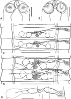
FIGURE 1. Hymenolepis bicauda n. sp. (A) Holotype, dorso-ventral view of scolex; (B) paratype, dorso-ventral view of scolex; (C) paratype, male mature proglottids; (D) paratype, hermaphroditic mature ... MoreFIGURE 1. Hymenolepis bicauda n. sp. (A) Holotype, dorso-ventral view of scolex; (B) paratype, dorso-ventral view of scolex; (C) paratype, male mature proglottids; (D) paratype, hermaphroditic mature proglottids; (E) paratype, genital ducts. Scale bars: A, B, E = 100 µm; C, D = 400 µm. |
Line Drawing 2
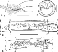
FIGURE 2. Hymenolepis bicauda n. sp. (A) Paratype, cirrus and vagina; (B) paratype, egg; (C) paratype, pregravid proglottid, showing uterus development; (D) paratype, gravid proglottid. Scale bars: A ... MoreFIGURE 2. Hymenolepis bicauda n. sp. (A) Paratype, cirrus and vagina; (B) paratype, egg; (C) paratype, pregravid proglottid, showing uterus development; (D) paratype, gravid proglottid. Scale bars: A = 50 µm; B = 20 µm; C, D = 400 µm. |
Photo Micrograph

FIGURE 3. Hymenolepis bicauda n. sp. Microphotographs of posterior ends of strobila in three paratypes showing terminal gravid proglottids splitting in the middle to form two tail-like structures. Not... MoreFIGURE 3. Hymenolepis bicauda n. sp. Microphotographs of posterior ends of strobila in three paratypes showing terminal gravid proglottids splitting in the middle to form two tail-like structures. Note eggs in the region of proglottid rupture. Scale bars: A, B, C = 500 µm. |
Scanning Electron Micrograph
|
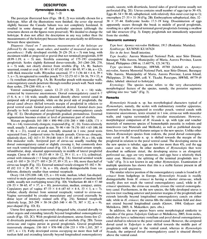
 FIGURE 1. Hymenolepis bicauda n. sp. (A) Holotype, dorso-ventral view of scolex; (B) paratype, dorso-ventral view of scolex; (C) paratype, male mature proglottids; (D) paratype, hermaphroditic mature proglottids; (E) paratype, genital ducts. Scale bars: A, B, E = 100 µm; C, D = 400 µm.
  FIGURE 2. Hymenolepis bicauda n. sp. (A) Paratype, cirrus and vagina; (B) paratype, egg; (C) paratype, pregravid proglottid, showing uterus development; (D) paratype, gravid proglottid. Scale bars: A = 50 µm; B = 20 µm; C, D = 400 µm.
 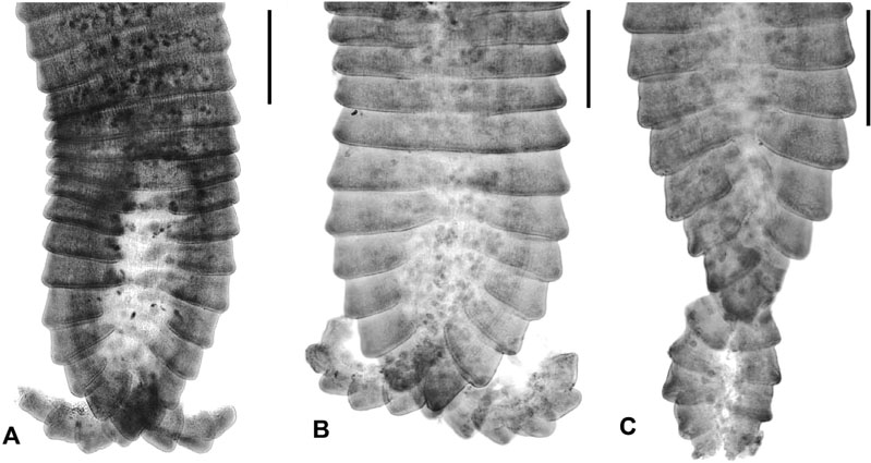 FIGURE 3. Hymenolepis bicauda n. sp. Microphotographs of posterior ends of strobila in three paratypes showing terminal gravid proglottids splitting in the middle to form two tail-like structures. Note eggs in the region of proglottid rupture. Scale bars: A, B, C = 500 µm.

