Line Drawing 1
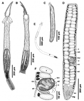
Fig. 7 Line drawings of Dollfusiella spinosa n. sp. from Pastinachus solocirostris Last, Manjaji & Yeatsley (BO-267) in the South
China Sea. A, scolex, lateral view; B, scolex, dorso-ventral view; C,... MoreFig. 7 Line drawings of Dollfusiella spinosa n. sp. from Pastinachus solocirostris Last, Manjaji & Yeatsley (BO-267) in the South
China Sea. A, scolex, lateral view; B, scolex, dorso-ventral view; C, bulb; D, mature segment; E, outline of complete specimen; F, detail of cirrus-sac, composite line drawing based on serial sections. Abbreviations: at, genital atrium, c, cirrus, cs, cirrus-sac, isv, internal seminal vesicle, mg, Mehlis gland, ov, ovary, t, testis, ut, uterus, vd, vas deferens, vit, vitelline follicle, vs, vaginal sphincter |
Line Drawing 2
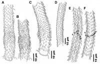
Fig. 8 Line drawings of the tentacular armature of Dollfusiella spinosa n. sp. from Pastinachus solocirostris Last, Manjaji & Yeatsley
(BO-267) in the South China Sea. A, basal tentacular armature, b... MoreFig. 8 Line drawings of the tentacular armature of Dollfusiella spinosa n. sp. from Pastinachus solocirostris Last, Manjaji & Yeatsley
(BO-267) in the South China Sea. A, basal tentacular armature, bothrial surface; B, basal tentacular armature, antibothrial surfacenote
opposing tentacular surface to tentacle illustrated in (A); C, basal tentacular armature, external surface; D, metabasal tentacular
armature, internal surface; E, metabasal tentacular armature, bothrial surface; F, metabasal tentacular armature, antibothrial surfacenote opposing tentacular surface to tentacle illustrated in (E) |
Photo Micrograph
|
Scanning Electron Micrograph

Fig. 9 Scanning electron micrographs of Dollfusiella spinosa n. sp. from Pastinachus solocirostris Last, Manjaji & Yeatsley (BO-267) in the South China Sea. A, complete specimen, dorso-ventral view; B... MoreFig. 9 Scanning electron micrographs of Dollfusiella spinosa n. sp. from Pastinachus solocirostris Last, Manjaji & Yeatsley (BO-267) in the South China Sea. A, complete specimen, dorso-ventral view; B, capilliform filitriches covering distal bothrial surface; C, palmate
spinitriches covering proximal bothrial surface; D, enlarged, palmate spinitriches covering scolex peduncle; E, anterior part of scolex,
lateral to dorso-ventral view; F, basal tentacular armature, bothrial surface; G, tentacular armature on basal swelling, antibothrial surface; H, metabasal tentacular armature, internal surface; I, basal tentacular armature, bothrial to external surface. Symbols: #, position on distal bothrial surface where microthrix micrograph (B) was taken; *, position on proximal bothrial surface where microthrix micrograph (C) was taken; ?, position on pars vaginalis where microthrix micrograph (D) was taken |
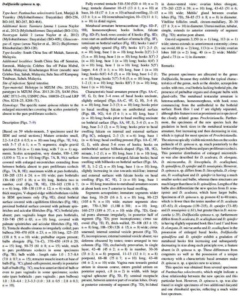
 Fig. 7 Line drawings of Dollfusiella spinosa n. sp. from Pastinachus solocirostris Last, Manjaji & Yeatsley (BO-267) in the South
China Sea. A, scolex, lateral view; B, scolex, dorso-ventral view; C, bulb; D, mature segment; E, outline of complete specimen; F, detail of cirrus-sac, composite line drawing based on serial sections. Abbreviations: at, genital atrium, c, cirrus, cs, cirrus-sac, isv, internal seminal vesicle, mg, Mehlis gland, ov, ovary, t, testis, ut, uterus, vd, vas deferens, vit, vitelline follicle, vs, vaginal sphincter
Fig. 7 Line drawings of Dollfusiella spinosa n. sp. from Pastinachus solocirostris Last, Manjaji & Yeatsley (BO-267) in the South
China Sea. A, scolex, lateral view; B, scolex, dorso-ventral view; C, bulb; D, mature segment; E, outline of complete specimen; F, detail of cirrus-sac, composite line drawing based on serial sections. Abbreviations: at, genital atrium, c, cirrus, cs, cirrus-sac, isv, internal seminal vesicle, mg, Mehlis gland, ov, ovary, t, testis, ut, uterus, vd, vas deferens, vit, vitelline follicle, vs, vaginal sphincter  Fig. 8 Line drawings of the tentacular armature of Dollfusiella spinosa n. sp. from Pastinachus solocirostris Last, Manjaji & Yeatsley
(BO-267) in the South China Sea. A, basal tentacular armature, bothrial surface; B, basal tentacular armature, antibothrial surfacenote
opposing tentacular surface to tentacle illustrated in (A); C, basal tentacular armature, external surface; D, metabasal tentacular
armature, internal surface; E, metabasal tentacular armature, bothrial surface; F, metabasal tentacular armature, antibothrial surfacenote opposing tentacular surface to tentacle illustrated in (E)
Fig. 8 Line drawings of the tentacular armature of Dollfusiella spinosa n. sp. from Pastinachus solocirostris Last, Manjaji & Yeatsley
(BO-267) in the South China Sea. A, basal tentacular armature, bothrial surface; B, basal tentacular armature, antibothrial surfacenote
opposing tentacular surface to tentacle illustrated in (A); C, basal tentacular armature, external surface; D, metabasal tentacular
armature, internal surface; E, metabasal tentacular armature, bothrial surface; F, metabasal tentacular armature, antibothrial surfacenote opposing tentacular surface to tentacle illustrated in (E) 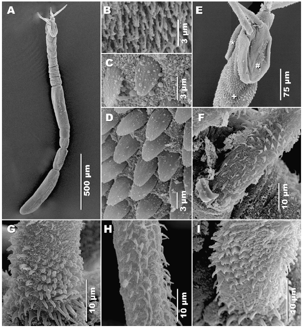 Fig. 9 Scanning electron micrographs of Dollfusiella spinosa n. sp. from Pastinachus solocirostris Last, Manjaji & Yeatsley (BO-267) in the South China Sea. A, complete specimen, dorso-ventral view; B, capilliform filitriches covering distal bothrial surface; C, palmate
spinitriches covering proximal bothrial surface; D, enlarged, palmate spinitriches covering scolex peduncle; E, anterior part of scolex,
lateral to dorso-ventral view; F, basal tentacular armature, bothrial surface; G, tentacular armature on basal swelling, antibothrial surface; H, metabasal tentacular armature, internal surface; I, basal tentacular armature, bothrial to external surface. Symbols: #, position on distal bothrial surface where microthrix micrograph (B) was taken; *, position on proximal bothrial surface where microthrix micrograph (C) was taken; ?, position on pars vaginalis where microthrix micrograph (D) was taken
Fig. 9 Scanning electron micrographs of Dollfusiella spinosa n. sp. from Pastinachus solocirostris Last, Manjaji & Yeatsley (BO-267) in the South China Sea. A, complete specimen, dorso-ventral view; B, capilliform filitriches covering distal bothrial surface; C, palmate
spinitriches covering proximal bothrial surface; D, enlarged, palmate spinitriches covering scolex peduncle; E, anterior part of scolex,
lateral to dorso-ventral view; F, basal tentacular armature, bothrial surface; G, tentacular armature on basal swelling, antibothrial surface; H, metabasal tentacular armature, internal surface; I, basal tentacular armature, bothrial to external surface. Symbols: #, position on distal bothrial surface where microthrix micrograph (B) was taken; *, position on proximal bothrial surface where microthrix micrograph (C) was taken; ?, position on pars vaginalis where microthrix micrograph (D) was taken 

