Line Drawing 1
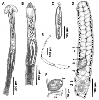
Fig. 4 Line drawings of Dollfusiella hemispinosa n. sp. from Himantura pastinacoides (Bleeker) (BO-12) in the South China Sea. A,
scolex, lateral view; B, scolex, dorso-ventral view; C, bulb; D, matu... MoreFig. 4 Line drawings of Dollfusiella hemispinosa n. sp. from Himantura pastinacoides (Bleeker) (BO-12) in the South China Sea. A,
scolex, lateral view; B, scolex, dorso-ventral view; C, bulb; D, mature segment; E, outline of entire cestode; F, detail of cirrus-sac, composite line drawing from serial sections. Abbreviations: c, cirrus, cs, cirrus-sac, isv, internal seminal vesicle, mg, Mehlis gland, ov,
ovary, t, testis, vit, vitelline follicle, vs, vaginal sphincter |
Line Drawing 2

Fig. 5 Line drawings of tentacular armature of Dollfusiella hemispinosa n. sp. from Himantura pastinacoides (Bleeker) (BO-12) in the South China Sea. A, basal tentacular armature, external surface; B,... MoreFig. 5 Line drawings of tentacular armature of Dollfusiella hemispinosa n. sp. from Himantura pastinacoides (Bleeker) (BO-12) in the South China Sea. A, basal tentacular armature, external surface; B, metabasal tentacular armature, antibothrial surface; C, metabasal tentacular armature, bothrial surfacenote opposing tentacular surface to tentacle illustrated in (B); D, basal tentacular armature, bothrial surface; E, basal tentacular armature, antibothrial surface |
Photo Micrograph
|
Scanning Electron Micrograph

Fig. 6 Scanning electron micrographs of Dollfusiella hemispinosa n. sp. from Himantura pastinacoides (Bleeker) (BO-12) in the South China Sea. A, scolex, lateral view; B, palmate
spinitriches coverin... MoreFig. 6 Scanning electron micrographs of Dollfusiella hemispinosa n. sp. from Himantura pastinacoides (Bleeker) (BO-12) in the South China Sea. A, scolex, lateral view; B, palmate
spinitriches covering proximal bothrial surface; C, enlarged, palmate spinitriches covering scolex peduncle; D, anterior part of scolex, dorso-ventral view; E, scolex, dorso-ventral view; F, anterior part of scolex, lateral view; G, metabasal tentacular armature, antibothrial surface; H, basal tentacular armature, bothrial surface; I, basal tentacular armature, antibothrial
surface; J, metabasal tentacular armature, external surface. Symbols:
*, position on proximal bothrial surface where microthrix micrograph (B) was taken; ?, position on pars vaginalis where microthrix micrograph (C) was taken |
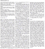
 Fig. 4 Line drawings of Dollfusiella hemispinosa n. sp. from Himantura pastinacoides (Bleeker) (BO-12) in the South China Sea. A,
scolex, lateral view; B, scolex, dorso-ventral view; C, bulb; D, mature segment; E, outline of entire cestode; F, detail of cirrus-sac, composite line drawing from serial sections. Abbreviations: c, cirrus, cs, cirrus-sac, isv, internal seminal vesicle, mg, Mehlis gland, ov,
ovary, t, testis, vit, vitelline follicle, vs, vaginal sphincter
Fig. 4 Line drawings of Dollfusiella hemispinosa n. sp. from Himantura pastinacoides (Bleeker) (BO-12) in the South China Sea. A,
scolex, lateral view; B, scolex, dorso-ventral view; C, bulb; D, mature segment; E, outline of entire cestode; F, detail of cirrus-sac, composite line drawing from serial sections. Abbreviations: c, cirrus, cs, cirrus-sac, isv, internal seminal vesicle, mg, Mehlis gland, ov,
ovary, t, testis, vit, vitelline follicle, vs, vaginal sphincter 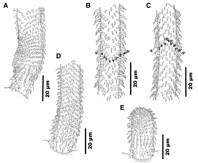 Fig. 5 Line drawings of tentacular armature of Dollfusiella hemispinosa n. sp. from Himantura pastinacoides (Bleeker) (BO-12) in the South China Sea. A, basal tentacular armature, external surface; B, metabasal tentacular armature, antibothrial surface; C, metabasal tentacular armature, bothrial surfacenote opposing tentacular surface to tentacle illustrated in (B); D, basal tentacular armature, bothrial surface; E, basal tentacular armature, antibothrial surface
 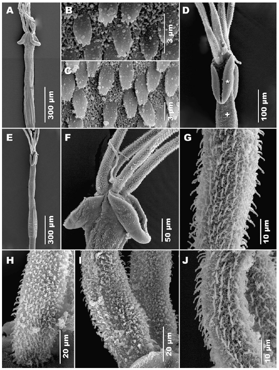 Fig. 6 Scanning electron micrographs of Dollfusiella hemispinosa n. sp. from Himantura pastinacoides (Bleeker) (BO-12) in the South China Sea. A, scolex, lateral view; B, palmate
spinitriches covering proximal bothrial surface; C, enlarged, palmate spinitriches covering scolex peduncle; D, anterior part of scolex, dorso-ventral view; E, scolex, dorso-ventral view; F, anterior part of scolex, lateral view; G, metabasal tentacular armature, antibothrial surface; H, basal tentacular armature, bothrial surface; I, basal tentacular armature, antibothrial
surface; J, metabasal tentacular armature, external surface. Symbols:
*, position on proximal bothrial surface where microthrix micrograph (B) was taken; ?, position on pars vaginalis where microthrix micrograph (C) was taken
Fig. 6 Scanning electron micrographs of Dollfusiella hemispinosa n. sp. from Himantura pastinacoides (Bleeker) (BO-12) in the South China Sea. A, scolex, lateral view; B, palmate
spinitriches covering proximal bothrial surface; C, enlarged, palmate spinitriches covering scolex peduncle; D, anterior part of scolex, dorso-ventral view; E, scolex, dorso-ventral view; F, anterior part of scolex, lateral view; G, metabasal tentacular armature, antibothrial surface; H, basal tentacular armature, bothrial surface; I, basal tentacular armature, antibothrial
surface; J, metabasal tentacular armature, external surface. Symbols:
*, position on proximal bothrial surface where microthrix micrograph (B) was taken; ?, position on pars vaginalis where microthrix micrograph (C) was taken 

