Line Drawing 1

Fig. 1 Line drawings of Dollfusiella angustiformis n. sp. from Himantura uarnacoides (Bleeker) (KA-146) in the Java Sea. A, scolex,
lateral view; B, scolex, dorso-ventral view; C, bulb; D, outline of... MoreFig. 1 Line drawings of Dollfusiella angustiformis n. sp. from Himantura uarnacoides (Bleeker) (KA-146) in the Java Sea. A, scolex,
lateral view; B, scolex, dorso-ventral view; C, bulb; D, outline of entire cestode; E, mature segment; F, detail of cirrus-sac, composite
line drawing from serial sections. Abbreviations: c, cirrus, cs, cirrus-sac, isv, internal seminal vesicle, mg, Mehlis gland, ov, ovary, t, testis, va, vagina, vit, vitelline follicle, vs, vaginal sphincter |
Line Drawing 2

Fig. 2 Line drawings of tentacular armature of Dollfusiella angustiformis n. sp. from Himantura uarnacoides (Bleeker) (KA-146) in the Java Sea. A, metabasal tentacular armature, antibothrial surface; ... MoreFig. 2 Line drawings of tentacular armature of Dollfusiella angustiformis n. sp. from Himantura uarnacoides (Bleeker) (KA-146) in the Java Sea. A, metabasal tentacular armature, antibothrial surface; B, metabasal tentacular armature, bothrial surfacenote opposing
tentacular surface to tentacle illustrated in (A); C, basal tentacular armature, external surface; D, basal tentacular armature, bothrial surface; E, basal tentacular armature, antibothrial surface |
Photo Micrograph
|
Scanning Electron Micrograph
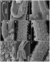
Fig. 3 Scanning electron micrographs of Dollfusiella angustiformis n. sp. from Himantura uarnacoides (Bleeker) (KA-146) in the Java Sea. A, complete specimen, lateral view; B, detailed view of bothriu... MoreFig. 3 Scanning electron micrographs of Dollfusiella angustiformis n. sp. from Himantura uarnacoides (Bleeker) (KA-146) in the Java Sea. A, complete specimen, lateral view; B, detailed view of bothriumnote enlarged, capilliform filitriches lining border between distal and proximal bothrial surfaces; C, basal
tentacular armature, bothrial surfaces; D, basal tentacular
armature, external surface; E, metabasal tentacular armature,
bothrial to external surface; F, metabasal tentacular armature,
antibothrial surface; G, palmate spinitriches covering proximal
bothrial surface; H, enlarged, palmate spinitriches covering
scolex peduncle; I, anterior part of scolex, lateral view; J, metabasal tentacular armature, external surface. Symbols: *, position on proximal bothrial surface where microthrix micrograph (G) was taken; ?, position on pars vaginalis where microthrix micrograph (H) was taken |
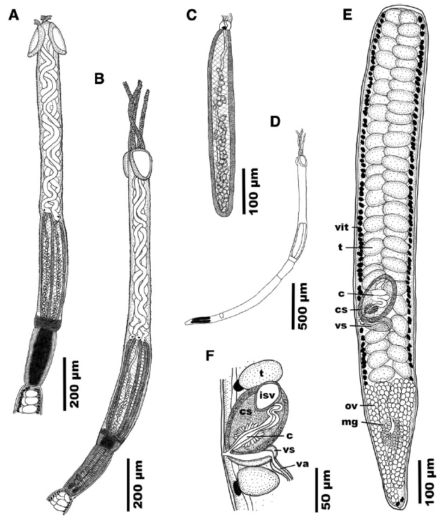 Fig. 1 Line drawings of Dollfusiella angustiformis n. sp. from Himantura uarnacoides (Bleeker) (KA-146) in the Java Sea. A, scolex,
lateral view; B, scolex, dorso-ventral view; C, bulb; D, outline of entire cestode; E, mature segment; F, detail of cirrus-sac, composite
line drawing from serial sections. Abbreviations: c, cirrus, cs, cirrus-sac, isv, internal seminal vesicle, mg, Mehlis gland, ov, ovary, t, testis, va, vagina, vit, vitelline follicle, vs, vaginal sphincter
Fig. 1 Line drawings of Dollfusiella angustiformis n. sp. from Himantura uarnacoides (Bleeker) (KA-146) in the Java Sea. A, scolex,
lateral view; B, scolex, dorso-ventral view; C, bulb; D, outline of entire cestode; E, mature segment; F, detail of cirrus-sac, composite
line drawing from serial sections. Abbreviations: c, cirrus, cs, cirrus-sac, isv, internal seminal vesicle, mg, Mehlis gland, ov, ovary, t, testis, va, vagina, vit, vitelline follicle, vs, vaginal sphincter 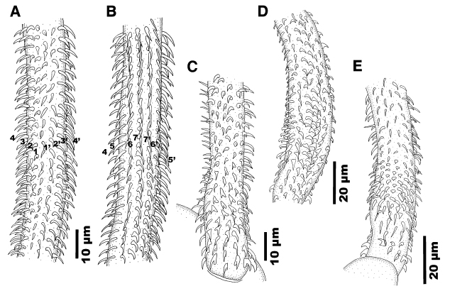 Fig. 2 Line drawings of tentacular armature of Dollfusiella angustiformis n. sp. from Himantura uarnacoides (Bleeker) (KA-146) in the Java Sea. A, metabasal tentacular armature, antibothrial surface; B, metabasal tentacular armature, bothrial surfacenote opposing
tentacular surface to tentacle illustrated in (A); C, basal tentacular armature, external surface; D, basal tentacular armature, bothrial surface; E, basal tentacular armature, antibothrial surface
Fig. 2 Line drawings of tentacular armature of Dollfusiella angustiformis n. sp. from Himantura uarnacoides (Bleeker) (KA-146) in the Java Sea. A, metabasal tentacular armature, antibothrial surface; B, metabasal tentacular armature, bothrial surfacenote opposing
tentacular surface to tentacle illustrated in (A); C, basal tentacular armature, external surface; D, basal tentacular armature, bothrial surface; E, basal tentacular armature, antibothrial surface  Fig. 3 Scanning electron micrographs of Dollfusiella angustiformis n. sp. from Himantura uarnacoides (Bleeker) (KA-146) in the Java Sea. A, complete specimen, lateral view; B, detailed view of bothriumnote enlarged, capilliform filitriches lining border between distal and proximal bothrial surfaces; C, basal
tentacular armature, bothrial surfaces; D, basal tentacular
armature, external surface; E, metabasal tentacular armature,
bothrial to external surface; F, metabasal tentacular armature,
antibothrial surface; G, palmate spinitriches covering proximal
bothrial surface; H, enlarged, palmate spinitriches covering
scolex peduncle; I, anterior part of scolex, lateral view; J, metabasal tentacular armature, external surface. Symbols: *, position on proximal bothrial surface where microthrix micrograph (G) was taken; ?, position on pars vaginalis where microthrix micrograph (H) was taken
Fig. 3 Scanning electron micrographs of Dollfusiella angustiformis n. sp. from Himantura uarnacoides (Bleeker) (KA-146) in the Java Sea. A, complete specimen, lateral view; B, detailed view of bothriumnote enlarged, capilliform filitriches lining border between distal and proximal bothrial surfaces; C, basal
tentacular armature, bothrial surfaces; D, basal tentacular
armature, external surface; E, metabasal tentacular armature,
bothrial to external surface; F, metabasal tentacular armature,
antibothrial surface; G, palmate spinitriches covering proximal
bothrial surface; H, enlarged, palmate spinitriches covering
scolex peduncle; I, anterior part of scolex, lateral view; J, metabasal tentacular armature, external surface. Symbols: *, position on proximal bothrial surface where microthrix micrograph (G) was taken; ?, position on pars vaginalis where microthrix micrograph (H) was taken 

