Line Drawing 1

Fig. 1 Line drawings of Poecilorhynchus perplexus n. g., n. sp. from Chiloscyllium punctatum from Nickol Bay, Western Australia. A, scolex, dorso-ventral view; B, scolex, lateral view; C, mature segme... MoreFig. 1 Line drawings of Poecilorhynchus perplexus n. g., n. sp. from Chiloscyllium punctatum from Nickol Bay, Western Australia. A, scolex, dorso-ventral view; B, scolex, lateral view; C, mature segment; D, bulb; E, detail of region of cirrus-sac. Abbreviations:c, cirrus,
cs cirrus-sac, ga, genital atrium, t, testis, ut, uterus, va, vagina, vit, vitelline follicle |
Line Drawing 2
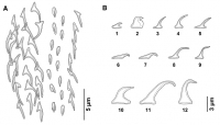
Fig. 2 Line drawings of the oncotaxy of Poecilorhynchus perplexus n. g., n. sp. from Chiloscyllium punctatum from Nickol Bay, Western Australia. A, schematic representation of the external surface of ... MoreFig. 2 Line drawings of the oncotaxy of Poecilorhynchus perplexus n. g., n. sp. from Chiloscyllium punctatum from Nickol Bay, Western Australia. A, schematic representation of the external surface of the mid-metabasal tentacular armaturenote space between
principle hook rows and two longitudinal files of hooks (chainettes); B, profiles of hooks: 1, uncinate hook of chainette, mid-metabasal region; 2, uncinate hook of chainette, distal metabasal region; 3, falcate principle hook with enlarged base, internal surface, basal to metabasal region; 4, 5, falcate principle hooks with enlarged bases, external surface, metabasal region; 6, 7, hooks 1(10), internal surface, basal region; 8, hooks 1(10), internal surface, metabasal region; 9, falcate principle hook with enlarged base and sinuous tip,
antibothrial surface, metabasal region; 10, enlarged uncinate hook, most proximal basal region; 11, falcate hook with recurved tip,
bothrial surface, basal region; 12, falcate hook with sinuous tip, bothrial surface, basal region |
Photo Micrograph
|
Scanning Electron Micrograph

Fig. 3 Scanning electron micrographs of Poecilorhynchus perplexus n. g., n. sp. from Chiloscyllium punctatum from Nickol Bay, Western Australia. A, scolex, lateral view; B, anterior part of scolex; C,... MoreFig. 3 Scanning electron micrographs of Poecilorhynchus perplexus n. g., n. sp. from Chiloscyllium punctatum from Nickol Bay, Western Australia. A, scolex, lateral view; B, anterior part of scolex; C, enlarged, palmate spinitriches covering scolex peduncle; D, detailed view of palmate spinithrix on scolex peduncle; E, smaller, palmate spinitriches covering proximal bothrial surface. Fig. 4 Scanning electron micrographs of Poecilorhynchus perplexus n. g., n. sp. from Chiloscyllium punctatum from Nickol Bay, Western Australia. A, basal tentacular armature, external surfacenote falcate hooks on bothrial surface (arrow); B, basal tentacular armature, antibothrial to external surfacenote uncinate hooks at base (arrow); C, basal tentacular armature, internal surface; D, transition from basal to metabasal tentacular armature, external surface; E, transition from basal to metabasal tentacular armature,
antibothrial to internal surfacenote falcate hooks with sinuous tip on antibothrial surface (arrow); F, transition from basal to metabasal tentacular armature, internal to antibothrial surface; G, metabasal tentacular armature, internal surface; H, metabasal
tentacular armature, external surfacenote falcate hooks with broadened base of principle hook rows and two longitudinal files of uncinate hooks forming chainette, separated from principle rows; I, detailed view of terminal hooks of principle rows (left) and two longitudinal files of hooks (right) on external surface of metabasal armaturenote space between principle hook rows and longitudinal files of hooks |
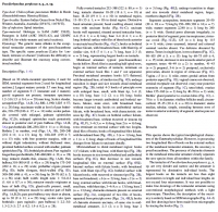
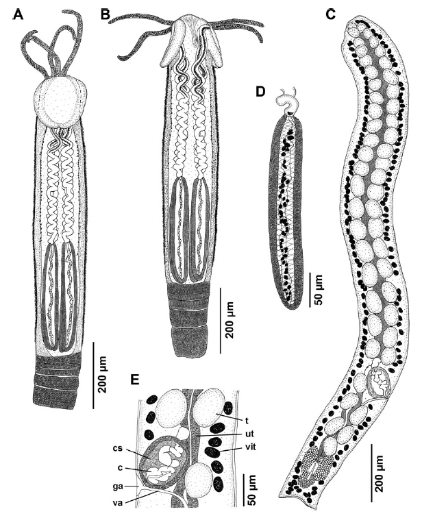 Fig. 1 Line drawings of Poecilorhynchus perplexus n. g., n. sp. from Chiloscyllium punctatum from Nickol Bay, Western Australia. A, scolex, dorso-ventral view; B, scolex, lateral view; C, mature segment; D, bulb; E, detail of region of cirrus-sac. Abbreviations:c, cirrus,
cs cirrus-sac, ga, genital atrium, t, testis, ut, uterus, va, vagina, vit, vitelline follicle
Fig. 1 Line drawings of Poecilorhynchus perplexus n. g., n. sp. from Chiloscyllium punctatum from Nickol Bay, Western Australia. A, scolex, dorso-ventral view; B, scolex, lateral view; C, mature segment; D, bulb; E, detail of region of cirrus-sac. Abbreviations:c, cirrus,
cs cirrus-sac, ga, genital atrium, t, testis, ut, uterus, va, vagina, vit, vitelline follicle 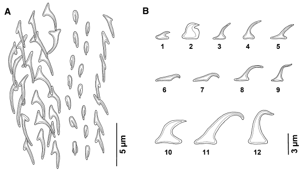 Fig. 2 Line drawings of the oncotaxy of Poecilorhynchus perplexus n. g., n. sp. from Chiloscyllium punctatum from Nickol Bay, Western Australia. A, schematic representation of the external surface of the mid-metabasal tentacular armaturenote space between
principle hook rows and two longitudinal files of hooks (chainettes); B, profiles of hooks: 1, uncinate hook of chainette, mid-metabasal region; 2, uncinate hook of chainette, distal metabasal region; 3, falcate principle hook with enlarged base, internal surface, basal to metabasal region; 4, 5, falcate principle hooks with enlarged bases, external surface, metabasal region; 6, 7, hooks 1(10), internal surface, basal region; 8, hooks 1(10), internal surface, metabasal region; 9, falcate principle hook with enlarged base and sinuous tip,
antibothrial surface, metabasal region; 10, enlarged uncinate hook, most proximal basal region; 11, falcate hook with recurved tip,
bothrial surface, basal region; 12, falcate hook with sinuous tip, bothrial surface, basal region
Fig. 2 Line drawings of the oncotaxy of Poecilorhynchus perplexus n. g., n. sp. from Chiloscyllium punctatum from Nickol Bay, Western Australia. A, schematic representation of the external surface of the mid-metabasal tentacular armaturenote space between
principle hook rows and two longitudinal files of hooks (chainettes); B, profiles of hooks: 1, uncinate hook of chainette, mid-metabasal region; 2, uncinate hook of chainette, distal metabasal region; 3, falcate principle hook with enlarged base, internal surface, basal to metabasal region; 4, 5, falcate principle hooks with enlarged bases, external surface, metabasal region; 6, 7, hooks 1(10), internal surface, basal region; 8, hooks 1(10), internal surface, metabasal region; 9, falcate principle hook with enlarged base and sinuous tip,
antibothrial surface, metabasal region; 10, enlarged uncinate hook, most proximal basal region; 11, falcate hook with recurved tip,
bothrial surface, basal region; 12, falcate hook with sinuous tip, bothrial surface, basal region 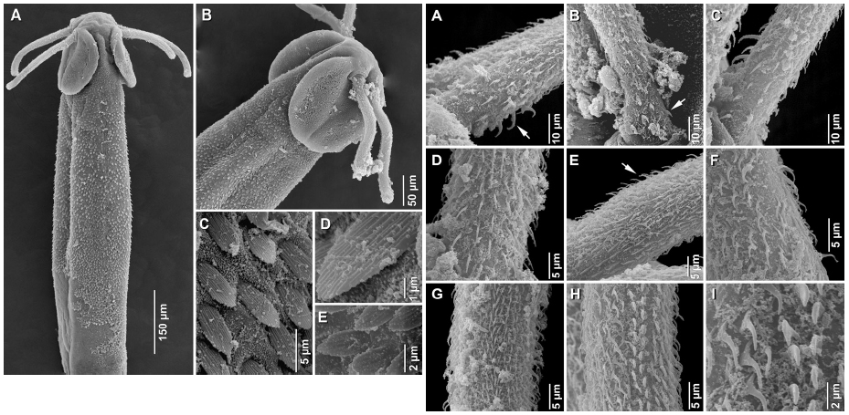 Fig. 3 Scanning electron micrographs of Poecilorhynchus perplexus n. g., n. sp. from Chiloscyllium punctatum from Nickol Bay, Western Australia. A, scolex, lateral view; B, anterior part of scolex; C, enlarged, palmate spinitriches covering scolex peduncle; D, detailed view of palmate spinithrix on scolex peduncle; E, smaller, palmate spinitriches covering proximal bothrial surface. Fig. 4 Scanning electron micrographs of Poecilorhynchus perplexus n. g., n. sp. from Chiloscyllium punctatum from Nickol Bay, Western Australia. A, basal tentacular armature, external surfacenote falcate hooks on bothrial surface (arrow); B, basal tentacular armature, antibothrial to external surfacenote uncinate hooks at base (arrow); C, basal tentacular armature, internal surface; D, transition from basal to metabasal tentacular armature, external surface; E, transition from basal to metabasal tentacular armature,
antibothrial to internal surfacenote falcate hooks with sinuous tip on antibothrial surface (arrow); F, transition from basal to metabasal tentacular armature, internal to antibothrial surface; G, metabasal tentacular armature, internal surface; H, metabasal
tentacular armature, external surfacenote falcate hooks with broadened base of principle hook rows and two longitudinal files of uncinate hooks forming chainette, separated from principle rows; I, detailed view of terminal hooks of principle rows (left) and two longitudinal files of hooks (right) on external surface of metabasal armaturenote space between principle hook rows and longitudinal files of hooks
Fig. 3 Scanning electron micrographs of Poecilorhynchus perplexus n. g., n. sp. from Chiloscyllium punctatum from Nickol Bay, Western Australia. A, scolex, lateral view; B, anterior part of scolex; C, enlarged, palmate spinitriches covering scolex peduncle; D, detailed view of palmate spinithrix on scolex peduncle; E, smaller, palmate spinitriches covering proximal bothrial surface. Fig. 4 Scanning electron micrographs of Poecilorhynchus perplexus n. g., n. sp. from Chiloscyllium punctatum from Nickol Bay, Western Australia. A, basal tentacular armature, external surfacenote falcate hooks on bothrial surface (arrow); B, basal tentacular armature, antibothrial to external surfacenote uncinate hooks at base (arrow); C, basal tentacular armature, internal surface; D, transition from basal to metabasal tentacular armature, external surface; E, transition from basal to metabasal tentacular armature,
antibothrial to internal surfacenote falcate hooks with sinuous tip on antibothrial surface (arrow); F, transition from basal to metabasal tentacular armature, internal to antibothrial surface; G, metabasal tentacular armature, internal surface; H, metabasal
tentacular armature, external surfacenote falcate hooks with broadened base of principle hook rows and two longitudinal files of uncinate hooks forming chainette, separated from principle rows; I, detailed view of terminal hooks of principle rows (left) and two longitudinal files of hooks (right) on external surface of metabasal armaturenote space between principle hook rows and longitudinal files of hooks 

