Line Drawing 1
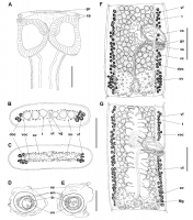
FIGURE 1. Harriscolex nathaliae n. sp. from Pseudoplatystoma corruscans. (A) Scolex, dorsoventral view (holotype MHNG-PLAT 82291). (B) Transverse cross section of mature proglottid at level of testes ... MoreFIGURE 1. Harriscolex nathaliae n. sp. from Pseudoplatystoma corruscans. (A) Scolex, dorsoventral view (holotype MHNG-PLAT 82291). (B) Transverse cross section of mature proglottid at level of testes posterior to cirrus sac (holotype MHNG-PLAT 82291). (C) Transverse cross section of mature proglottid at the level of the ovary (holotype MHNG-PLAT 82291). (D, E) Detail of eggs (MHNG-PLAT 22917, host field No. Py 5076). (F) Mature proglottid, dorsal view (paratype MHNG-PLAT 82290, field number Ar 1667). (G) Gravid proglottid, ventral view (holotype MHNG-PLAT 82291). Abbreviations: cp, cone-shaped projection; cs, cirrus sac; doc, dorsal osmoregulatory canal; em, embryophore; ga, genital atrium; gc, gland cells;
lh, larval hooks; lm, longitudinal musculature; Mg, Mehliss gland; oe, outer envelope; on, oncosphere; ov, ovary; t, testes; ut, uterus; vc, vaginal canal; vf, vitelline follicles; voc, ventral osmoregulatory canal; vs, vaginal sphincter. Scale bars: A, F, G = 250 µm; B, C = 200 µm; D, E = 20 µm. |
Line Drawing 2
|
Photo Micrograph
|
Scanning Electron Micrograph
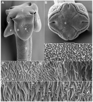
FIGURE 2. Scanning electron micrographs of Harriscolex nathaliae n. sp. (A) Latero-dorsoventral view of scolex; letters indicate location of high-magnification sections shown in CI. (B) Apical view o... MoreFIGURE 2. Scanning electron micrographs of Harriscolex nathaliae n. sp. (A) Latero-dorsoventral view of scolex; letters indicate location of high-magnification sections shown in CI. (B) Apical view of scolex. (C) Apical surface of scolex. (D) Marginal surface of sucker. (E) Luminal surface of sucker. (F) Nonadherent surface of sucker. (G) Proliferation zone surface. (H) Immature proglottid surface. (I) Mature proglottid surface. Filled black arrow shows cone-shaped projections; filled white arrow shows aciculate filitriches; empty white arrow shows small gladiate spinitriches; dashed arrow shows large gladiate spinitriches. Scale bars: A, B = 100 µm; CI = 1 µm. |
 FIGURE 1. Harriscolex nathaliae n. sp. from Pseudoplatystoma corruscans. (A) Scolex, dorsoventral view (holotype MHNG-PLAT 82291). (B) Transverse cross section of mature proglottid at level of testes posterior to cirrus sac (holotype MHNG-PLAT 82291). (C) Transverse cross section of mature proglottid at the level of the ovary (holotype MHNG-PLAT 82291). (D, E) Detail of eggs (MHNG-PLAT 22917, host field No. Py 5076). (F) Mature proglottid, dorsal view (paratype MHNG-PLAT 82290, field number Ar 1667). (G) Gravid proglottid, ventral view (holotype MHNG-PLAT 82291). Abbreviations: cp, cone-shaped projection; cs, cirrus sac; doc, dorsal osmoregulatory canal; em, embryophore; ga, genital atrium; gc, gland cells;
lh, larval hooks; lm, longitudinal musculature; Mg, Mehliss gland; oe, outer envelope; on, oncosphere; ov, ovary; t, testes; ut, uterus; vc, vaginal canal; vf, vitelline follicles; voc, ventral osmoregulatory canal; vs, vaginal sphincter. Scale bars: A, F, G = 250 µm; B, C = 200 µm; D, E = 20 µm.
FIGURE 1. Harriscolex nathaliae n. sp. from Pseudoplatystoma corruscans. (A) Scolex, dorsoventral view (holotype MHNG-PLAT 82291). (B) Transverse cross section of mature proglottid at level of testes posterior to cirrus sac (holotype MHNG-PLAT 82291). (C) Transverse cross section of mature proglottid at the level of the ovary (holotype MHNG-PLAT 82291). (D, E) Detail of eggs (MHNG-PLAT 22917, host field No. Py 5076). (F) Mature proglottid, dorsal view (paratype MHNG-PLAT 82290, field number Ar 1667). (G) Gravid proglottid, ventral view (holotype MHNG-PLAT 82291). Abbreviations: cp, cone-shaped projection; cs, cirrus sac; doc, dorsal osmoregulatory canal; em, embryophore; ga, genital atrium; gc, gland cells;
lh, larval hooks; lm, longitudinal musculature; Mg, Mehliss gland; oe, outer envelope; on, oncosphere; ov, ovary; t, testes; ut, uterus; vc, vaginal canal; vf, vitelline follicles; voc, ventral osmoregulatory canal; vs, vaginal sphincter. Scale bars: A, F, G = 250 µm; B, C = 200 µm; D, E = 20 µm.  FIGURE 2. Scanning electron micrographs of Harriscolex nathaliae n. sp. (A) Latero-dorsoventral view of scolex; letters indicate location of high-magnification sections shown in CI. (B) Apical view of scolex. (C) Apical surface of scolex. (D) Marginal surface of sucker. (E) Luminal surface of sucker. (F) Nonadherent surface of sucker. (G) Proliferation zone surface. (H) Immature proglottid surface. (I) Mature proglottid surface. Filled black arrow shows cone-shaped projections; filled white arrow shows aciculate filitriches; empty white arrow shows small gladiate spinitriches; dashed arrow shows large gladiate spinitriches. Scale bars: A, B = 100 µm; CI = 1 µm.

