Cestode Scientific Name
| Species ID | 12754 |
|---|---|
| Order | Diphyllidea |
| Family | |
| Subfamily | |
| Genus | Halysioncum |
| Species | raschii |
| Authority | (Campbell & Andrade, 1997) Caira, Marques, Jensen, Kuchta & Ivanov, 2013 |
| Taxonomic Status | Valid |
| Valid Name | |
| Synonyms | |
| Genus Record | No |
| Type Species | No |
| Verified | No |
| Verified By | V. Ivanov, R. Kuchta |
| Citation(s) |
Campbell, R. A. and M. Andrade. 1997. Echinobothrium raschii n. sp. (Cestoda: Diphyllidea) from Rhinoraja longi (Chondrichthyes, Rajoidei) in the Bering Sea. Journal of Parasitology 83: 115-120. (115) Download PDFCaira, J. N., F. P. L. Marques, K. Jensen, R. Kuchta, and V. Ivanov. 2013. Phylogenetic analysis and reconfiguration of genera in the cestode order Diphyllidea. International Journal for Parasitology 43: 621-639. (6204) Download PDF |
| Redescription | |
| Scientific Name Notes |
Record Data
| Date (MM/DD/YYYY) | Action | User Name |
|---|---|---|
| 10/18/2013 | Created | B. Barbeau |
| 08/20/2014 | Modified | |
| 02/03/2016 | Modified | K. Mojica |
| 02/03/2016 | Modified | J. Caira |
| 03/08/2016 | Modified | J. Caira |
| 03/23/2016 | Modified | B. Barbeau |
| 05/04/2016 | Modified | B. Barbeau |
| 08/23/2016 | Modified | R. Kuchta |
Type Host
| Host Class | Chondrichthyes | ||||||
|---|---|---|---|---|---|---|---|
| Host Order | Rajiformes | ||||||
| Host Family | Arhynchobatidae | ||||||
|
Type Host (Literal) |
|
||||||
|
Type Host (Valid) |
|
||||||
| Additional Host(s) | |||||||
| Site in Host | spiral intestine | ||||||
| Host Notes |
Type Locality
| Country | U.S.A. |
|---|---|
| Body of Water | Bering Sea |
| Island(s) | |
| City/Region | Alaska |
| Coordinates | |
| DD Latitude | 56° 07.6'N |
| DD Longitude | 168° 20.6'W |
| Additional Localities | |
| Locality Notes | at 100-103 fathoms |
Specimens
| Type Material | Holotype: USNPC no. 86767. Paratypes: USNPC no. 86768-86770 (3 specimens); BMNH no. 1996.7.26.3-6 (3 specimens). |
|---|---|
| Total Number of Type Specimens | based on 70 specimens |
| Voucher Material | |
| Specimen Notes |
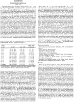
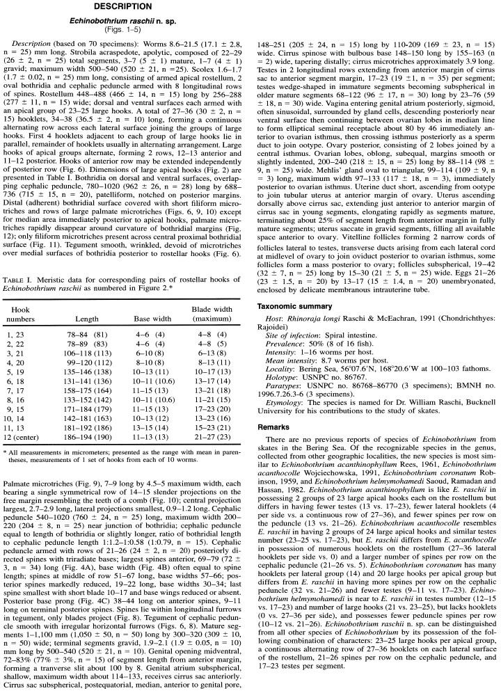
Data are given as in original description unless otherwise indicated.

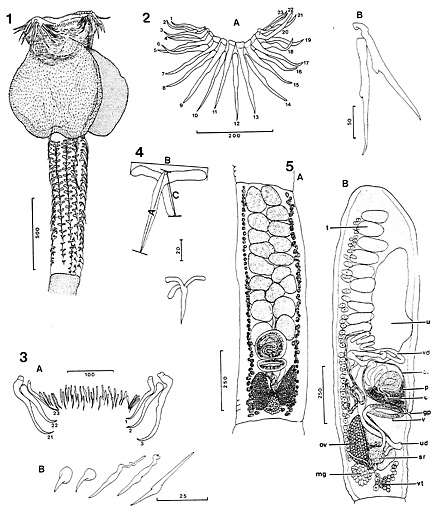 FIGURES 1-5. Echinobothrium raschii n. sp. from Bering Sea skate 1. Scolex, paratype. 2. Rostellar hooks, apical view of large hooks (A) detail of large hooks (B). 3. Rostellar hooklets, continuous row between apical groups of large hooks (A); variation in hooklet form (B). 4. First (anterior) and last (posterior) hooks from row on cephalic peduncle. Interlandmark distances for length (A) and width (B) and posterior extension (C) measurements indicated by brackets on first hook. 5. Proglottid anatomy. (A) Mature proglottid in ventral view; (B) gravid proglottid in lateral view showing ovary in section. (Abreviations: c, cirrus; cs, cirrus sac; gp, gential pore; mg, Mehlis gland; ov, ovary; p, prostate cells; sr, seminal recepticle; t, testis; u, uterus; ud, uterine duct; v, vagina; vd, vas deferens; vt, vitelline follicles.
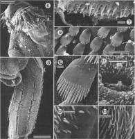
 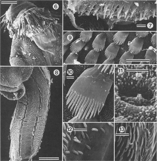 FIGURES 6-13. Scanning electron micrographs of E. raschii. All figures oriented with anterior end toward top of page. 6. Scolex, note large apical hooks of rostellum and microtriches on distal (adherent) surface of bothridium. 7. Hooklets forming contiguous row between large apical groups of hooks. 8. Cephalic peduncle. Note that only tips of spines protrude from longitudinal furrows. 9. Detail of rows of palmate microtriches on median distal bothridial surface. 10. Detail of palmate microthrix bearing 15 spinous processes; note small filamentous microtriches covering bothridial surface. 11. Detail of filamentous microtriches and circular pit on proximal bothridial surface. 12. Transition from proximal (right) to distal (left) surfaces of bothridium along posterior bothridial margin; note absence of palmate microtriches on proximal surface (right). 13. Detail of distal bothridial surface on lateral margin showing palmate microtriches; porelike openings are visible where microtriches were attached. Scale bars Figures 5 and 8, 100 µm; scale bars in Figures 7, 9, 12, and 13, 10 µm; scale bar in Figures 10, 11, 1 µm.
Best viewed in Firefox


