Line Drawing 1
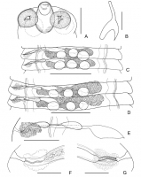
Fig. 1 Sawadalepis prima n. g., n. sp. Holotype. A, dorsoventral view of scolex; B, rostellar hook; C, male mature proglottides; D, hermaphroditic mature proglottides; E, genital ducts; F, cirrus; G, ... MoreFig. 1 Sawadalepis prima n. g., n. sp. Holotype. A, dorsoventral view of scolex; B, rostellar hook; C, male mature proglottides; D, hermaphroditic mature proglottides; E, genital ducts; F, cirrus; G, vagina. Scale-bars: A, 250 µm; B, 10 µm; C, D, 500 µm; E, 200 µm; F, G, 100 µm |
Line Drawing 2
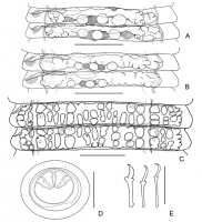
Fig. 2 Sawadalepis prima n. g., n. sp. Holotype. A, postmature proglottides, dorsal view showing uterine development; B, terminal postmature proglottides, dorsal view showing immature uterus crossing ... MoreFig. 2 Sawadalepis prima n. g., n. sp. Holotype. A, postmature proglottides, dorsal view showing uterine development; B, terminal postmature proglottides, dorsal view showing immature uterus crossing osmoregulatory canals; C, gravid proglottides, dorsal view showing saccate uterus with dorsal uterine diverticula; D, egg; E, embryonic hooks. Scale-bars: A-C, 500 µm; D, 30 µm; E, 10 µm |
Photo Micrograph

Fig. 3 Sawadalepis prima n. g., n. sp. Holotype. A, egg; B, C, rostellar hooks in lateral view (B) and view
from anterior surface showing narrow hook guard (C). Scale-bars: A, 20 µm; B, C, 10 µm |
Scanning Electron Micrograph
|
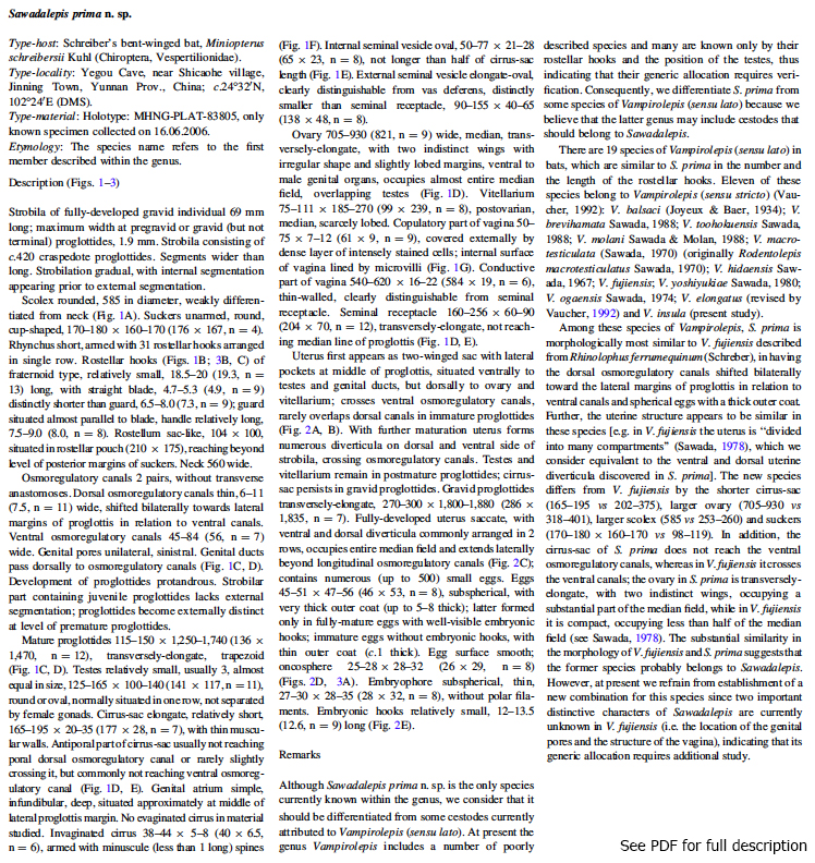
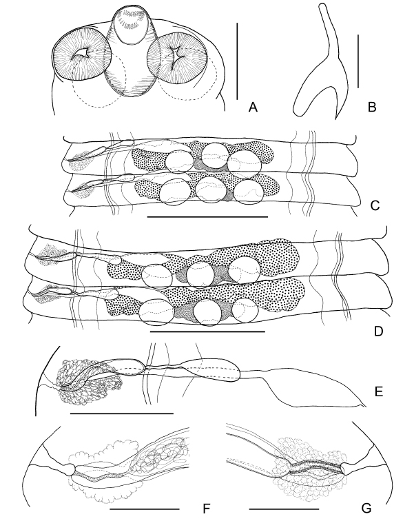 Fig. 1 Sawadalepis prima n. g., n. sp. Holotype. A, dorsoventral view of scolex; B, rostellar hook; C, male mature proglottides; D, hermaphroditic mature proglottides; E, genital ducts; F, cirrus; G, vagina. Scale-bars: A, 250 µm; B, 10 µm; C, D, 500 µm; E, 200 µm; F, G, 100 µm
 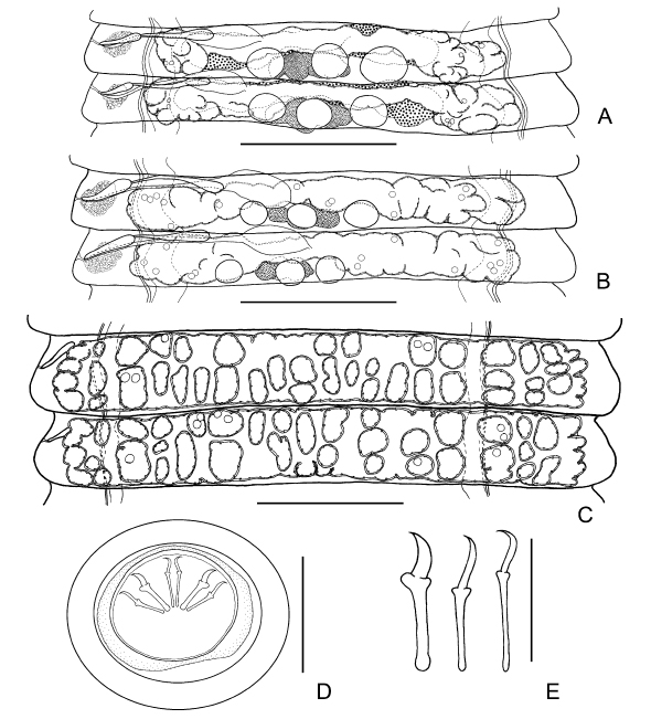 Fig. 2 Sawadalepis prima n. g., n. sp. Holotype. A, postmature proglottides, dorsal view showing uterine development; B, terminal postmature proglottides, dorsal view showing immature uterus crossing osmoregulatory canals; C, gravid proglottides, dorsal view showing saccate uterus with dorsal uterine diverticula; D, egg; E, embryonic hooks. Scale-bars: A-C, 500 µm; D, 30 µm; E, 10 µm
 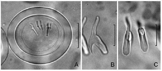 Fig. 3 Sawadalepis prima n. g., n. sp. Holotype. A, egg; B, C, rostellar hooks in lateral view (B) and view
from anterior surface showing narrow hook guard (C). Scale-bars: A, 20 µm; B, C, 10 µm
Fig. 3 Sawadalepis prima n. g., n. sp. Holotype. A, egg; B, C, rostellar hooks in lateral view (B) and view
from anterior surface showing narrow hook guard (C). Scale-bars: A, 20 µm; B, C, 10 µm 

