Cestode Scientific Name
| Species ID | 12723 |
|---|---|
| Order | Cyclophyllidea |
| Family | Taeniidae |
| Subfamily | |
| Genus | Urocystidium |
| Species | gemmiparum |
| Authority | Beddard, 1912 |
| Taxonomic Status | Synonym |
| Valid Name | Hydatigera taeniaeformis (Batsch, 1786) Lamarck, 1816 |
| Synonyms | |
| Genus Record | No |
| Type Species | Yes |
| Verified | Yes |
| Verified By | V. K. Haukisalmi |
| Citation(s) |
Beddard, F. E. 1912. Contributions on the anatomy and systematic arrangement of the Cestoidea. VI. On an asexual tapeworm from the rodent, Fiber zibethicus, showing a new form of asexual propagation, and on the supposed sexual form. Proceedings of the Zoological Society of London 1912(2): 822-850. (6154) Download PDF |
| Redescription | |
| Scientific Name Notes | According to Adam (1964), Urocystidium gemmiparum is a synonym of Taenia taeniaeformis Batsch, 1786. |
Record Data
| Date (MM/DD/YYYY) | Action | User Name |
|---|---|---|
| 10/07/2013 | Created | B. Barbeau, V. K. Haukisalmi |
| 01/16/2015 | Modified | |
| 05/04/2021 | Modified | V. Haukisalmi |
Type Host
| Host Class | Mammalia | ||||||
|---|---|---|---|---|---|---|---|
| Host Order | Rodentia | ||||||
| Host Family | Cricetidae | ||||||
|
Type Host (Literal) |
|
||||||
|
Type Host (Valid) |
|
||||||
| Additional Host(s) | |||||||
| Site in Host | liver | ||||||
| Host Notes |
Type Locality
| Country | |
|---|---|
| Body of Water | |
| Island(s) | |
| City/Region | |
| Coordinates | |
| DD Latitude | |
| DD Longitude | |
| Additional Localities | |
| Locality Notes |
Specimens
| Type Material | |
|---|---|
| Total Number of Type Specimens | |
| Voucher Material | |
| Specimen Notes |
Data are given as in original description unless otherwise indicated.
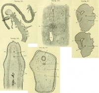
 Text-fig. 113. The upper represents the entire asexual form of Urocystidium gemmiparum enlarged by about one-third. The lower figure is the posterior end of the same individual more magnified. For explanation see text. Text-fig. 114. Part of a transverse section through a proglottid of the asexual form of Urocystidium gemmiparum. A, spaces in the middle of proglottid with darkly staining walls referred to in text. l.m. Longitudinal muscular layer of cortex, within which is seen delicate transverse layer of muscles. n.s. Nerve-cord. v.d. Dorsal excretory tube. v.v. Ventral excretory tube. Text-fig. 115. A more highly magnified section through excretory vessels and adjacent structures of the asexual form of Urocystidium gemmiparum. d.v. Dorsal vessels surrounded by circular muscle-fibre. m. Layer of transverse muscle-fibres separating the cortex (to the right) from the medulla (to the left). n. One of several tubes forming the excretory network and lying, as is shown, in the cortex as well as in the medulla. t.v. Transverse commissural vessel forking to join the ventral vessel (v.v.), which is also bifurcate to receive the branches. Text-fig. 116. Transverse section through middle of strobila of oldest bud of the asexual form of Urocystidium gemmiparum. l.v. excretory tubes. n. Nerve-cord. x. Tube of doubtful significance described in text. Text-fig. 117. Two consecutive sections from another region of the same bud as that which is represented in text-figure 116, to illustrate the opening of the tube x on to the exterior. In the upper figure the orifice (o.) on to the exterior is shown and the commencement of the invagination leading towards the tube x. In the lower figure the outgrowth of the tube x to meet the invagination is seen. n. Nerve-cord. l.v. Water vascular tubes.
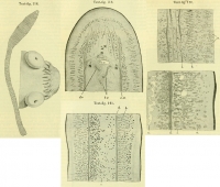
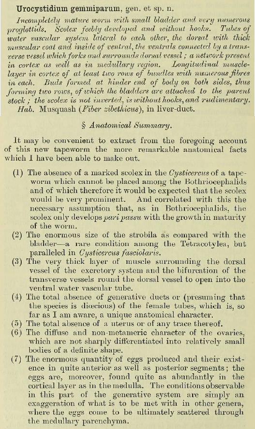
 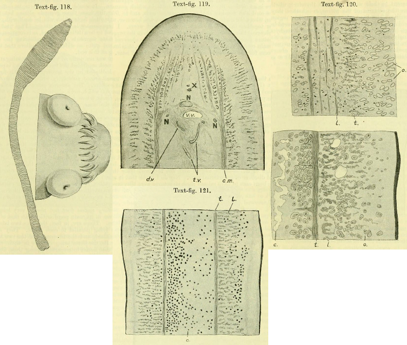 Text-fig. 118. The sexual form of Urocystidium gemmiparum (?). The left-hand figure represents the entire worm magnified about twice; the right-hand figure represents the scoelx with a double crown of hooks more highly magnified. Text-fig. 119. Part of a transverse section through a proglottid of the sexual worm. c.m. Circular muscles. d.v. Dorsal vessel of the water vascular system. N. The three laterally running nerve-cords. t.v. Transverse water vascular trunk. v.v. Ventral trunk. X. Large longitudinal muscular fibres referred to in the text as frequently running within a space. In the neighborhood of the water vascular trunks delicate muscle fibres are shown running chiefly in a dorso-ventral direction, which may be associated with the dilation and contraction of the water vascular tubes. Text-fig. 120. Two sections through portions of proglottids of the sexual worm. The upper figure is a longitudinal section in the more anterior part of the body, showing the smaller ova lying among and to the inside of the longitudinal muscles (l.); these smaller ova lie in what is probably generative tissue, not clearly indicated in the drawing. t. Transverse muscle fibres. o. Mature ova scattered through medulla and also at the opposite side of the figure in the cortex. The lower figure represents a transverse section through a more posteriorly situated segment. c. A cavity which may possibly represent a uterus; two spaces are shown in the cortex which may or may not belong in the same category. l. Longitudinal muscles. o. Ripe ova showing a nucleus. t. Transverse muscles. Text-fig. 121. Part of a transverse section through a proglottid of the sexually mature worm in which the ripe ova (o.) have been very deeply stained and are seen to be scattered through the cortex as well as the medulla. l. Longitudinal muscles. t. Tranverse muscles.
 Best viewed in Firefox


