Line Drawing 1
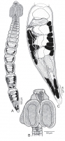
Fig. 1. Line drawings of Anteropora species. AC, A. joannae, new species. A, whole worm, holotype (MZUM[P] 2013.7[H]); B, scolex, paratype (USNPC 106528); C, terminal proglottid, paratype (USNPC 1065... MoreFig. 1. Line drawings of Anteropora species. AC, A. joannae, new species. A, whole worm, holotype (MZUM[P] 2013.7[H]); B, scolex, paratype (USNPC 106528); C, terminal proglottid, paratype (USNPC 106528). |
Line Drawing 2
|
Photo Micrograph
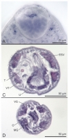
Fig. 8. Light micrographs of Anteropora species. A, primarily glandular apical organ of A. joannae, new species, holotype (MZUM[P] 2013.7[H]); C, crosssection through proglottid of A. joannae, new spe... MoreFig. 8. Light micrographs of Anteropora species. A, primarily glandular apical organ of A. joannae, new species, holotype (MZUM[P] 2013.7[H]); C, crosssection through proglottid of A. joannae, new species anterior to cirrus-sac, paratype (USNPC 106526); D, cross-section through proglottid of A. joannae, new species at level of ovary, paratype (USNPC 106526). (Abbreviations: ESV, external seminal vesicle; MG, Mehlis gland; O, ovary; T, testis; U, uterus; VG, vagina; VT, vitelline follicle.) |
Scanning Electron Micrograph
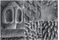
Fig. 2. Scanning electron micrographs of Anteropora species. AF, A. joannae, new species. A, scolex (small letters indicate location of
details in BF); B, surface of apical modifi cation of scolex ... MoreFig. 2. Scanning electron micrographs of Anteropora species. AF, A. joannae, new species. A, scolex (small letters indicate location of
details in BF); B, surface of apical modifi cation of scolex proper; C, surface of scolex proper at base of apical modifi cation; D, distal
bothridial surface; E, proximal bothridial surface; F, surface of posterior margin of proglottid. |
2013_7(H).jpg)
2013.7(H)_BO127_1HTsc.jpg)
2013.7(H)_BO127_1HTtp.jpg)
2013.7(H)_BO127_1HTao.jpg)
2013.7(H)_BO127_1HTww.jpg)
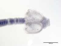
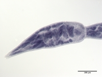
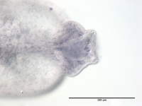
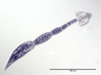
 Fig. 1. Line drawings of Anteropora species. AC, A. joannae, new species. A, whole worm, holotype (MZUM[P] 2013.7[H]); B, scolex, paratype (USNPC 106528); C, terminal proglottid, paratype (USNPC 106528).
 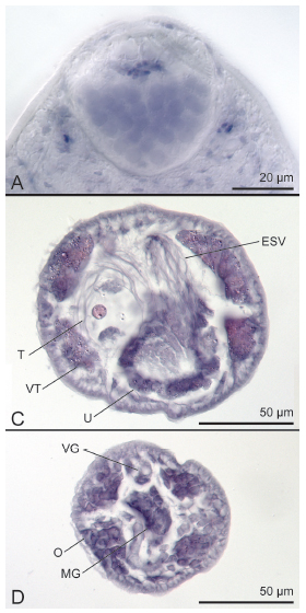 Fig. 8. Light micrographs of Anteropora species. A, primarily glandular apical organ of A. joannae, new species, holotype (MZUM[P] 2013.7[H]); C, crosssection through proglottid of A. joannae, new species anterior to cirrus-sac, paratype (USNPC 106526); D, cross-section through proglottid of A. joannae, new species at level of ovary, paratype (USNPC 106526). (Abbreviations: ESV, external seminal vesicle; MG, Mehlis gland; O, ovary; T, testis; U, uterus; VG, vagina; VT, vitelline follicle.)
 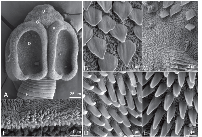 Fig. 2. Scanning electron micrographs of Anteropora species. AF, A. joannae, new species. A, scolex (small letters indicate location of
details in BF); B, surface of apical modifi cation of scolex proper; C, surface of scolex proper at base of apical modifi cation; D, distal
bothridial surface; E, proximal bothridial surface; F, surface of posterior margin of proglottid.
Fig. 2. Scanning electron micrographs of Anteropora species. AF, A. joannae, new species. A, scolex (small letters indicate location of
details in BF); B, surface of apical modifi cation of scolex proper; C, surface of scolex proper at base of apical modifi cation; D, distal
bothridial surface; E, proximal bothridial surface; F, surface of posterior margin of proglottid. 2013_7(H).jpg) MZUM(P) 2013.7(H), holotype slide
MZUM(P) 2013.7(H), holotype slide 2013.7(H)_BO127_1HTsc.jpg) MZUM(P) 2013.7(H), scolex of holotype
MZUM(P) 2013.7(H), scolex of holotype 2013.7(H)_BO127_1HTtp.jpg) MZUM(P) 2013.7(H), terminal proglottid of holotype
MZUM(P) 2013.7(H), terminal proglottid of holotype 2013.7(H)_BO127_1HTao.jpg) MZUM(P) 2013.7(H), apical organ of holotype
MZUM(P) 2013.7(H), apical organ of holotype 2013.7(H)_BO127_1HTww.jpg) MZUM(P) 2013.7(H), holotype, whole worm
MZUM(P) 2013.7(H), holotype, whole worm  USNPC No. 106528, paratype slide
 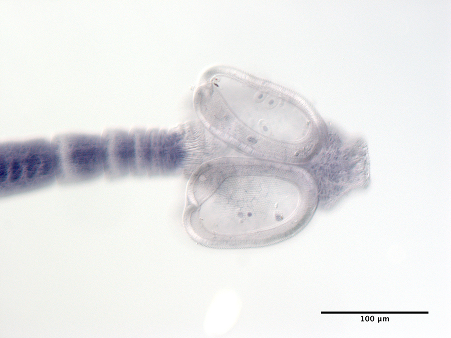 USNPC No. 106528, scolex of paratype
 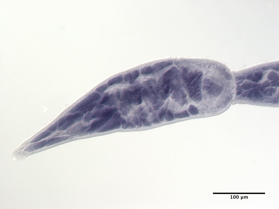 USNPC No. 106528, terminal proglottid of paratype
 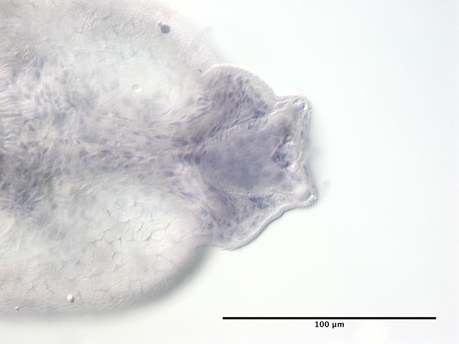 USNPC No. 106528, apical organ of paratype
 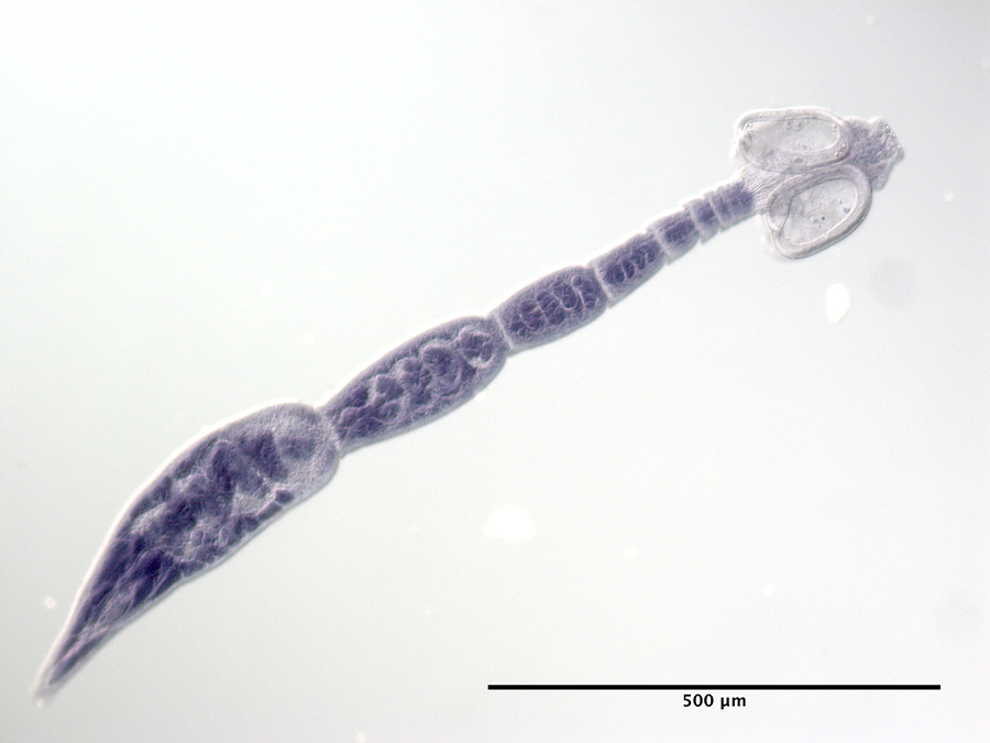 USNPC No. 106528, paratype, whole worm

