Line Drawing 1
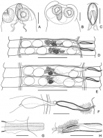
Fig. 1. Hymenolepis apodemi sp. nov. A holotype, dorsoventral view of scolex; B - paratype 18.31.4.30, dorsoventral view of retracted scolex; C holotype, rostellar pouch; D paratype 18.31.4.30, ... MoreFig. 1. Hymenolepis apodemi sp. nov. A holotype, dorsoventral view of scolex; B - paratype 18.31.4.30, dorsoventral view of retracted scolex; C holotype, rostellar pouch; D paratype 18.31.4.30, male mature proglottids; E holotype, hermaphroditic mature proglottids; F holotype, genital ducts; G holotype, cirrus; H vagina. Scale bars: A, B, C, F, H = 100 μm; D, E = 500 μm; G = 20 μm |
Line Drawing 2

Fig. 2. Hymenolepis apodemi sp. nov. A holotype, postmature proglottids from dorsal side, showing uterus development; B paratype 18.31.4.30, pregravid proglottids from ventral side, showing appear... MoreFig. 2. Hymenolepis apodemi sp. nov. A holotype, postmature proglottids from dorsal side, showing uterus development; B paratype 18.31.4.30, pregravid proglottids from ventral side, showing appearance of dorsal uterine diverticula; C voucher 18.31.4.70, cross-section showing dorsal and ventral uterine diverticula; D paratype 18.31.4.30, gravid proglottid from dorsal side, showing saccate uterus with dorsal uterine diverticula; E paratype 18.31.4.30, egg; F paratype 18.31.4.30, embryonic hooks. Scale bars: A, B, C, D = 500 μm; E = 25 μm; F = 15 μm |
Photo Micrograph
|
Scanning Electron Micrograph
|


 Fig. 1. Hymenolepis apodemi sp. nov. A holotype, dorsoventral view of scolex; B - paratype 18.31.4.30, dorsoventral view of retracted scolex; C holotype, rostellar pouch; D paratype 18.31.4.30, male mature proglottids; E holotype, hermaphroditic mature proglottids; F holotype, genital ducts; G holotype, cirrus; H vagina. Scale bars: A, B, C, F, H = 100 μm; D, E = 500 μm; G = 20 μm
  Fig. 2. Hymenolepis apodemi sp. nov. A holotype, postmature proglottids from dorsal side, showing uterus development; B paratype 18.31.4.30, pregravid proglottids from ventral side, showing appearance of dorsal uterine diverticula; C voucher 18.31.4.70, cross-section showing dorsal and ventral uterine diverticula; D paratype 18.31.4.30, gravid proglottid from dorsal side, showing saccate uterus with dorsal uterine diverticula; E paratype 18.31.4.30, egg; F paratype 18.31.4.30, embryonic hooks. Scale bars: A, B, C, D = 500 μm; E = 25 μm; F = 15 μm

