Line Drawing 1
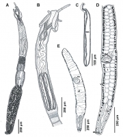
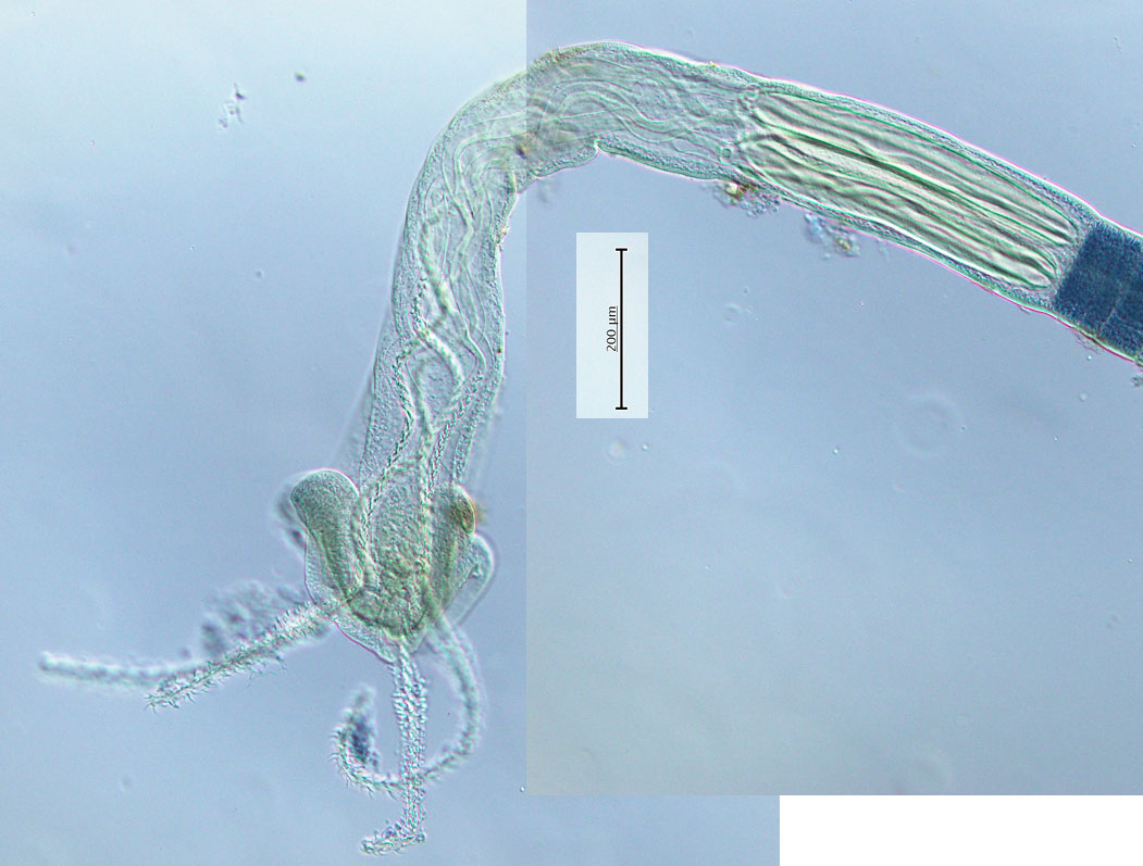
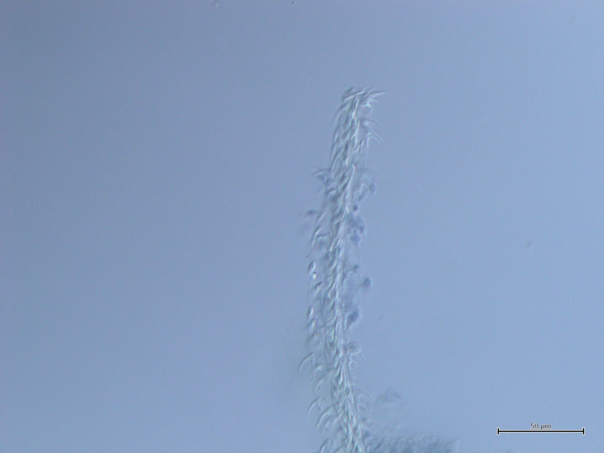
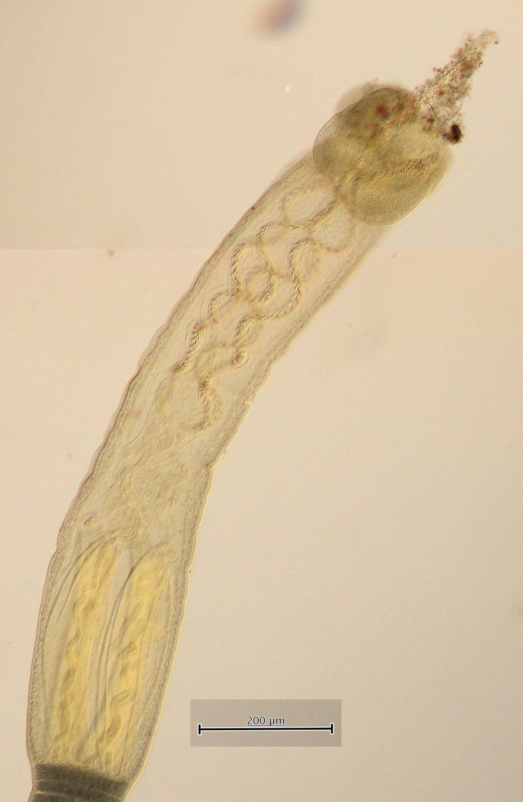
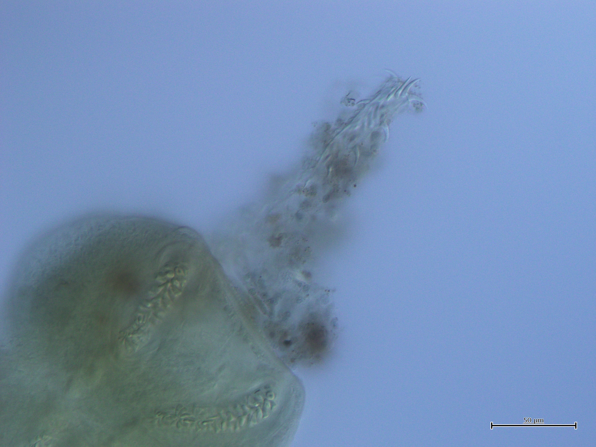
FIGURE 4. Prochristianella jensenae n. sp. from Pastinachus solocirostris A. Complete specimen. B. Scolex. C. Bulb; note absence of gland-cells. D. Mature segment. E. Gravid segment. Abbreviations: cs... MoreFIGURE 4. Prochristianella jensenae n. sp. from Pastinachus solocirostris A. Complete specimen. B. Scolex. C. Bulb; note absence of gland-cells. D. Mature segment. E. Gravid segment. Abbreviations: cs, cirrus sac; isv, internal seminal vesicle; mg, Mehlis' gland; ov, ovary; rm, retractor muscle; t, testis; ut, uterus; vit, vitelline follicle. |
Line Drawing 2
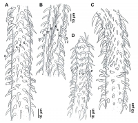
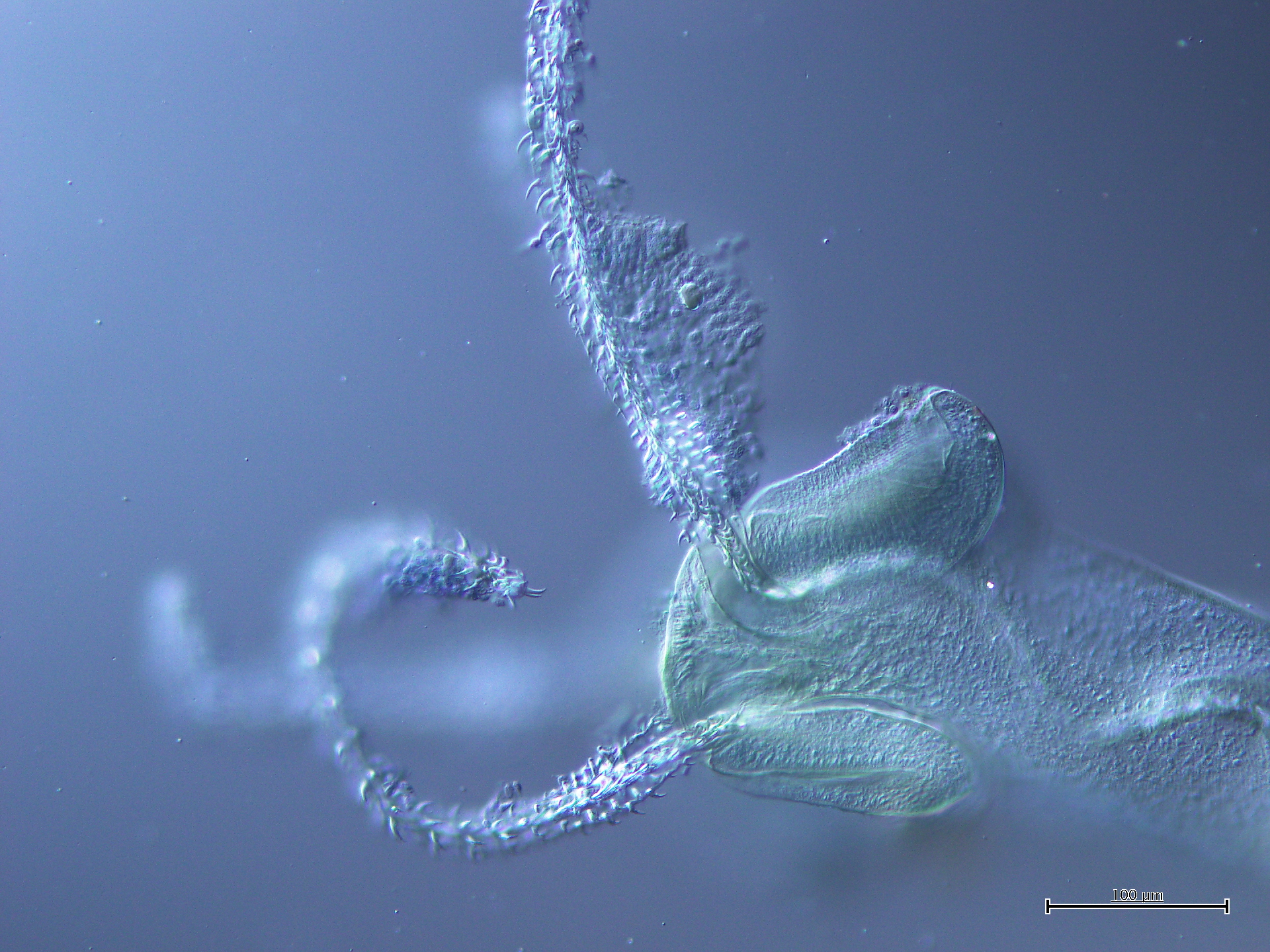
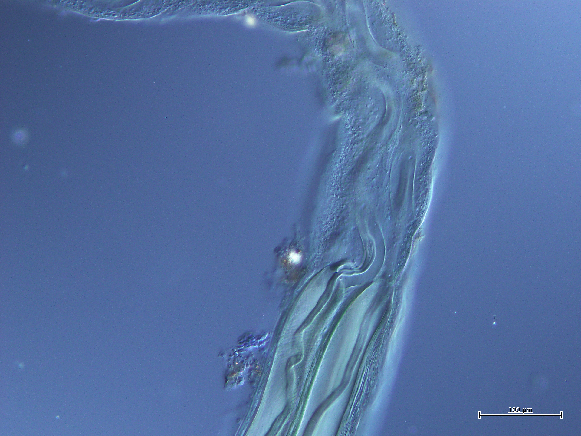
FIGURE 5. Prochristianella jensenae n. sp. from Pastinachus gracilicaudus (A, D) and P. solocirostris (B, C). Tentacular armature. A. Basal and metabasal regions, internal surface. B. Metabasal region... MoreFIGURE 5. Prochristianella jensenae n. sp. from Pastinachus gracilicaudus (A, D) and P. solocirostris (B, C). Tentacular armature. A. Basal and metabasal regions, internal surface. B. Metabasal region, antibothrial surface. C. Basal and metabasal regions, antibothrial surface. D. Basal and metabasal regions, bothrial surface. |
Photo Micrograph
|
Scanning Electron Micrograph
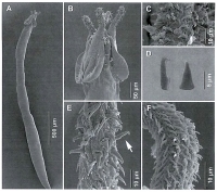
FIGURE 6. Prochristianella jensenae n. sp. from Pastinachus solcirostris. Scanning electron micrographs. A. Complete specimen. B. Scolex in lateral view. C. Tentacular armature; metabasal region, both... MoreFIGURE 6. Prochristianella jensenae n. sp. from Pastinachus solcirostris. Scanning electron micrographs. A. Complete specimen. B. Scolex in lateral view. C. Tentacular armature; metabasal region, bothrial surface. D. Billhooks in lateral (left) and dorso-ventral (right) views. E. Tentacular armature, basal region, internal surface; note erect billhook. F. Tentacular armature, metabasal region, external surface. |


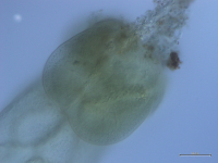
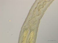

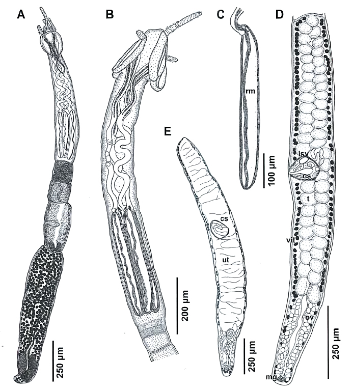 FIGURE 4. Prochristianella jensenae n. sp. from Pastinachus solocirostris A. Complete specimen. B. Scolex. C. Bulb; note absence of gland-cells. D. Mature segment. E. Gravid segment. Abbreviations: cs, cirrus sac; isv, internal seminal vesicle; mg, Mehlis' gland; ov, ovary; rm, retractor muscle; t, testis; ut, uterus; vit, vitelline follicle.
 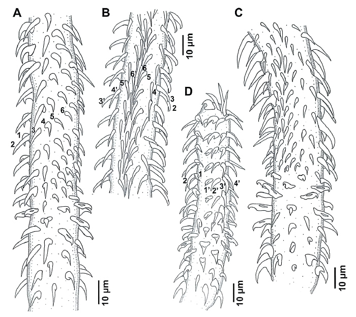 FIGURE 5. Prochristianella jensenae n. sp. from Pastinachus gracilicaudus (A, D) and P. solocirostris (B, C). Tentacular armature. A. Basal and metabasal regions, internal surface. B. Metabasal region, antibothrial surface. C. Basal and metabasal regions, antibothrial surface. D. Basal and metabasal regions, bothrial surface.
 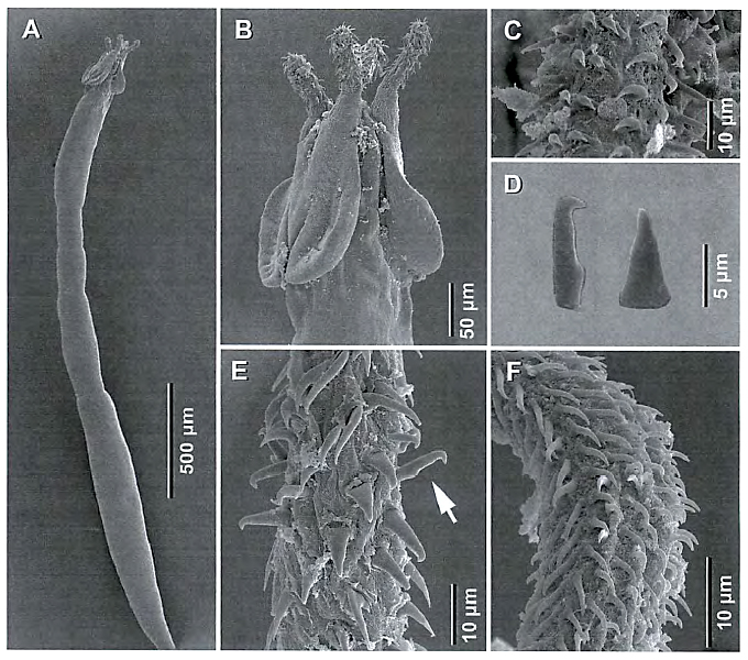 FIGURE 6. Prochristianella jensenae n. sp. from Pastinachus solcirostris. Scanning electron micrographs. A. Complete specimen. B. Scolex in lateral view. C. Tentacular armature; metabasal region, bothrial surface. D. Billhooks in lateral (left) and dorso-ventral (right) views. E. Tentacular armature, basal region, internal surface; note erect billhook. F. Tentacular armature, metabasal region, external surface.