Line Drawing 1

FIGURES 3639. Echinobothrium weipaense n. sp. line drawings. (36) Entire worm, scale bar = 50 µm. (37) Scolex, scale bar = 15 µm. (38) Apical armature, dorsoventral view, 16 apical hooks type A, 1'... MoreFIGURES 3639. Echinobothrium weipaense n. sp. line drawings. (36) Entire worm, scale bar = 50 µm. (37) Scolex, scale bar = 15 µm. (38) Apical armature, dorsoventral view, 16 apical hooks type A, 1'5' apical hooks type B, scale bar = 10 µm. (39) Mature proglottid, dorsal view, scale bar = 30 µm. |
Line Drawing 2
|
Photo Micrograph
|
Scanning Electron Micrograph
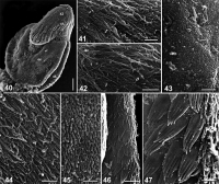
FIGURES 4047. Echinobothrium weipaense n. sp., scanning electron micrographs. (40) Scolex proper, scale bar = 10 µm. Small numbers indicate locations of details shown in Figures 4142, 44, 46. (41) D... MoreFIGURES 4047. Echinobothrium weipaense n. sp., scanning electron micrographs. (40) Scolex proper, scale bar = 10 µm. Small numbers indicate locations of details shown in Figures 4142, 44, 46. (41) Detail of hooklets, scale bar = 2.5 µm. (42) Apex of scolex surface, scale bar = 2 µm.
(43) Cephalic peduncle surface, scale bar = 1 µm. (44) Distal bothrial surface, scale bar = 1 µm. (45) Immature proglottid surface, scale bar = 0.8 µm. (46) Proximal bothrial surface, scale bar = 2 µm. (47) Detail of microtriches on proximal bothrial surface, scale bar = 1 µm. |
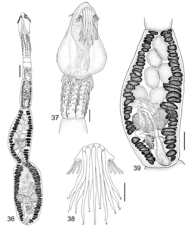 FIGURES 3639. Echinobothrium weipaense n. sp. line drawings. (36) Entire worm, scale bar = 50 µm. (37) Scolex, scale bar = 15 µm. (38) Apical armature, dorsoventral view, 16 apical hooks type A, 1'5' apical hooks type B, scale bar = 10 µm. (39) Mature proglottid, dorsal view, scale bar = 30 µm.
 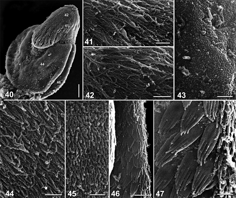 FIGURES 4047. Echinobothrium weipaense n. sp., scanning electron micrographs. (40) Scolex proper, scale bar = 10 µm. Small numbers indicate locations of details shown in Figures 4142, 44, 46. (41) Detail of hooklets, scale bar = 2.5 µm. (42) Apex of scolex surface, scale bar = 2 µm.
(43) Cephalic peduncle surface, scale bar = 1 µm. (44) Distal bothrial surface, scale bar = 1 µm. (45) Immature proglottid surface, scale bar = 0.8 µm. (46) Proximal bothrial surface, scale bar = 2 µm. (47) Detail of microtriches on proximal bothrial surface, scale bar = 1 µm.
FIGURES 4047. Echinobothrium weipaense n. sp., scanning electron micrographs. (40) Scolex proper, scale bar = 10 µm. Small numbers indicate locations of details shown in Figures 4142, 44, 46. (41) Detail of hooklets, scale bar = 2.5 µm. (42) Apex of scolex surface, scale bar = 2 µm.
(43) Cephalic peduncle surface, scale bar = 1 µm. (44) Distal bothrial surface, scale bar = 1 µm. (45) Immature proglottid surface, scale bar = 0.8 µm. (46) Proximal bothrial surface, scale bar = 2 µm. (47) Detail of microtriches on proximal bothrial surface, scale bar = 1 µm. 

