Line Drawing 1
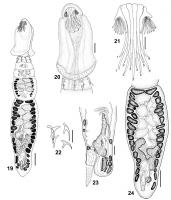
FIGURES 1924. Echinobothrium sematanense n. sp., line drawings. (19) Entire worm, scale bar = 50 µm. (20) Scolex, scale bar = 20 µm. (21) Apical armature, dorsoventral view, 16 type-A hooks, 1'5' t... MoreFIGURES 1924. Echinobothrium sematanense n. sp., line drawings. (19) Entire worm, scale bar = 50 µm. (20) Scolex, scale bar = 20 µm. (21) Apical armature, dorsoventral view, 16 type-A hooks, 1'5' type-B hooks, scale bar = 10 µm. (22) Detail of spines on the cephalic peduncle, scale bar = 10 µm. (23) Detail of terminal genitalia, lateral view, scale bar = 25 µm. (24) Gravid proglottid, ventral view, scale bar = 50 µm. |
Line Drawing 2
|
Photo Micrograph
|
Scanning Electron Micrograph
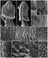
FIGURES 2535. Echinobothrium sematanense n. sp., scanning electron micrographs. (25) Scolex, dorsoventral view, scale bar = 20 µm. Small numbers
indicate locations of details shown in Figures 2931,... MoreFIGURES 2535. Echinobothrium sematanense n. sp., scanning electron micrographs. (25) Scolex, dorsoventral view, scale bar = 20 µm. Small numbers
indicate locations of details shown in Figures 2931, 3334. (26) Scolex, lateral view, scale bar = 20 µm. Small numbers indicate locations of details shown in Figures 27, 28, 32, 35. (27) Detail of hooklets, scale bar = 3 µm. (28) Apex of scolex surface, scale bar = 1.5 µm. (29) Central region of distal bothrial surface, scale bar = 1 µm. (30) Lateral and posterior regions of distal bothrial surface, scale bar = 1 µm. (31) Surface of tissue covering hooks, scale bar = 1 µm. (32) Proximal bothrial surface, anterior regions, scale bar = 1 µm. (33) Proximal bothrial surface, posterior region, scale bar = 1 µm. (34) Cephalic peduncle surface, scale bar = 1 µm. (35) Surface of immature proglottid, scale bar = 1 µm. |
 FIGURES 1924. Echinobothrium sematanense n. sp., line drawings. (19) Entire worm, scale bar = 50 µm. (20) Scolex, scale bar = 20 µm. (21) Apical armature, dorsoventral view, 16 type-A hooks, 1'5' type-B hooks, scale bar = 10 µm. (22) Detail of spines on the cephalic peduncle, scale bar = 10 µm. (23) Detail of terminal genitalia, lateral view, scale bar = 25 µm. (24) Gravid proglottid, ventral view, scale bar = 50 µm.
  FIGURES 2535. Echinobothrium sematanense n. sp., scanning electron micrographs. (25) Scolex, dorsoventral view, scale bar = 20 µm. Small numbers
indicate locations of details shown in Figures 2931, 3334. (26) Scolex, lateral view, scale bar = 20 µm. Small numbers indicate locations of details shown in Figures 27, 28, 32, 35. (27) Detail of hooklets, scale bar = 3 µm. (28) Apex of scolex surface, scale bar = 1.5 µm. (29) Central region of distal bothrial surface, scale bar = 1 µm. (30) Lateral and posterior regions of distal bothrial surface, scale bar = 1 µm. (31) Surface of tissue covering hooks, scale bar = 1 µm. (32) Proximal bothrial surface, anterior regions, scale bar = 1 µm. (33) Proximal bothrial surface, posterior region, scale bar = 1 µm. (34) Cephalic peduncle surface, scale bar = 1 µm. (35) Surface of immature proglottid, scale bar = 1 µm.
FIGURES 2535. Echinobothrium sematanense n. sp., scanning electron micrographs. (25) Scolex, dorsoventral view, scale bar = 20 µm. Small numbers
indicate locations of details shown in Figures 2931, 3334. (26) Scolex, lateral view, scale bar = 20 µm. Small numbers indicate locations of details shown in Figures 27, 28, 32, 35. (27) Detail of hooklets, scale bar = 3 µm. (28) Apex of scolex surface, scale bar = 1.5 µm. (29) Central region of distal bothrial surface, scale bar = 1 µm. (30) Lateral and posterior regions of distal bothrial surface, scale bar = 1 µm. (31) Surface of tissue covering hooks, scale bar = 1 µm. (32) Proximal bothrial surface, anterior regions, scale bar = 1 µm. (33) Proximal bothrial surface, posterior region, scale bar = 1 µm. (34) Cephalic peduncle surface, scale bar = 1 µm. (35) Surface of immature proglottid, scale bar = 1 µm. 

