Line Drawing 1
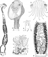
FIGURES 16. Echinobothrium tetabuanense n. sp., line drawings. (1) Entire worm in ventral view, scale bar = 200 µm. (2) Scolex, scale bar = 50 µm. (3) Apical armature, dorsoventral view, 16 type-A h... MoreFIGURES 16. Echinobothrium tetabuanense n. sp., line drawings. (1) Entire worm in ventral view, scale bar = 200 µm. (2) Scolex, scale bar = 50 µm. (3) Apical armature, dorsoventral view, 16 type-A hooks, 1'5' type-B hooks, scale bar = 25 µm. (4) Detail of spines on the cephalic peduncle: (A) Anteriormost spines, (B) posteriormost spines, scale bar = 22 µm. (5) Detail of terminal genitalia in dorsal view, scale bar = 35 µm. (6) Gravid proglottid
in ventral view, scale bar = 50 µm. Abbreviations: cs, cirrus sac; gp, genital pore; isv, internal seminal vesicle; mg, Mehlis gland; ov, ovary; ud, uterine duct; vd, vas deferens; vf, vitelline follicle; vg, vagina. |
Line Drawing 2
|
Photo Micrograph
|
Scanning Electron Micrograph

FIGURES 718. Echinobothrium tetabuanense n. sp., scanning electron micrographs. (7) Lateral view of scolex, scale bar = 100 µm. Small numbers
indicate locations of details shown in Figures 14, 16, a... MoreFIGURES 718. Echinobothrium tetabuanense n. sp., scanning electron micrographs. (7) Lateral view of scolex, scale bar = 100 µm. Small numbers
indicate locations of details shown in Figures 14, 16, and 17. (8) Scolex proper, dorsoventral view, scale bar = 50 µm. Small numbers indicate locations
of details shown in Figures 10, 12, 13, and 15. (9) Detail of hooklets, scale bar = 10 µm. Small number indicates location of detail shown in Figure 11.
(10) Surface of apex of scolex, scale bar = 2 µm. (11) Surface of tissue covering hooklets, scale bar = 1 µm. (12) Central region of distal bothrial surface,
scale bar = 1 µm. (13) Lateral and posterior regions of distal bothrial surface, scale bar = 1.2 µm. (14) Lateral surface of scolex proper, scale bar = 1.5 µm. (15) Border between distal and proximal bothrial surfaces, scale bar = 2 µm. (16) Proximal bothrial surface, scale bar = 2 µm. (17) Cephalic
peduncle surface, scale bar = 1 µm. (18) Mature proglottid surface, scale bar = 1.3 µm. |
 FIGURES 16. Echinobothrium tetabuanense n. sp., line drawings. (1) Entire worm in ventral view, scale bar = 200 µm. (2) Scolex, scale bar = 50 µm. (3) Apical armature, dorsoventral view, 16 type-A hooks, 1'5' type-B hooks, scale bar = 25 µm. (4) Detail of spines on the cephalic peduncle: (A) Anteriormost spines, (B) posteriormost spines, scale bar = 22 µm. (5) Detail of terminal genitalia in dorsal view, scale bar = 35 µm. (6) Gravid proglottid
in ventral view, scale bar = 50 µm. Abbreviations: cs, cirrus sac; gp, genital pore; isv, internal seminal vesicle; mg, Mehlis gland; ov, ovary; ud, uterine duct; vd, vas deferens; vf, vitelline follicle; vg, vagina.
FIGURES 16. Echinobothrium tetabuanense n. sp., line drawings. (1) Entire worm in ventral view, scale bar = 200 µm. (2) Scolex, scale bar = 50 µm. (3) Apical armature, dorsoventral view, 16 type-A hooks, 1'5' type-B hooks, scale bar = 25 µm. (4) Detail of spines on the cephalic peduncle: (A) Anteriormost spines, (B) posteriormost spines, scale bar = 22 µm. (5) Detail of terminal genitalia in dorsal view, scale bar = 35 µm. (6) Gravid proglottid
in ventral view, scale bar = 50 µm. Abbreviations: cs, cirrus sac; gp, genital pore; isv, internal seminal vesicle; mg, Mehlis gland; ov, ovary; ud, uterine duct; vd, vas deferens; vf, vitelline follicle; vg, vagina. 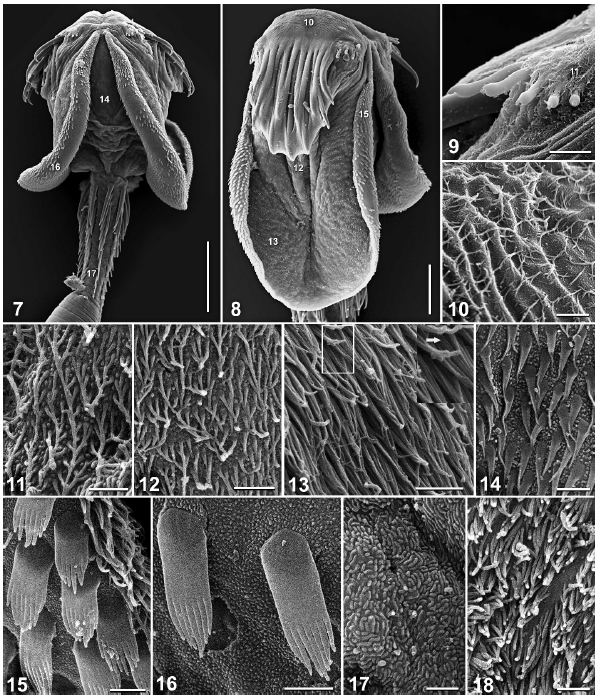 FIGURES 718. Echinobothrium tetabuanense n. sp., scanning electron micrographs. (7) Lateral view of scolex, scale bar = 100 µm. Small numbers
indicate locations of details shown in Figures 14, 16, and 17. (8) Scolex proper, dorsoventral view, scale bar = 50 µm. Small numbers indicate locations
of details shown in Figures 10, 12, 13, and 15. (9) Detail of hooklets, scale bar = 10 µm. Small number indicates location of detail shown in Figure 11.
(10) Surface of apex of scolex, scale bar = 2 µm. (11) Surface of tissue covering hooklets, scale bar = 1 µm. (12) Central region of distal bothrial surface,
scale bar = 1 µm. (13) Lateral and posterior regions of distal bothrial surface, scale bar = 1.2 µm. (14) Lateral surface of scolex proper, scale bar = 1.5 µm. (15) Border between distal and proximal bothrial surfaces, scale bar = 2 µm. (16) Proximal bothrial surface, scale bar = 2 µm. (17) Cephalic
peduncle surface, scale bar = 1 µm. (18) Mature proglottid surface, scale bar = 1.3 µm.
FIGURES 718. Echinobothrium tetabuanense n. sp., scanning electron micrographs. (7) Lateral view of scolex, scale bar = 100 µm. Small numbers
indicate locations of details shown in Figures 14, 16, and 17. (8) Scolex proper, dorsoventral view, scale bar = 50 µm. Small numbers indicate locations
of details shown in Figures 10, 12, 13, and 15. (9) Detail of hooklets, scale bar = 10 µm. Small number indicates location of detail shown in Figure 11.
(10) Surface of apex of scolex, scale bar = 2 µm. (11) Surface of tissue covering hooklets, scale bar = 1 µm. (12) Central region of distal bothrial surface,
scale bar = 1 µm. (13) Lateral and posterior regions of distal bothrial surface, scale bar = 1.2 µm. (14) Lateral surface of scolex proper, scale bar = 1.5 µm. (15) Border between distal and proximal bothrial surfaces, scale bar = 2 µm. (16) Proximal bothrial surface, scale bar = 2 µm. (17) Cephalic
peduncle surface, scale bar = 1 µm. (18) Mature proglottid surface, scale bar = 1.3 µm. 

