Line Drawing 1
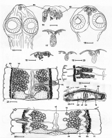
FIGURES 14-24. Proteocephalus synodontis Woodland, 1925. 14, Holotype (BMNH 1961.4.10.87-102), Khartoum, Sudan,
'&:olex, dorsoventral view. IS, 17-20. Apical organs. IS. Holotype, Khartoum. 17. From ... MoreFIGURES 14-24. Proteocephalus synodontis Woodland, 1925. 14, Holotype (BMNH 1961.4.10.87-102), Khartoum, Sudan,
'&:olex, dorsoventral view. IS, 17-20. Apical organs. IS. Holotype, Khartoum. 17. From Synodontis schall, Girba, Sudan; 18.
Immature tapeworm from Synodontis schall, Lake Turkana, Kenya; 19. From Synodontis schall, Kostl, Sudan; 20. From Synodontis
caudovittata, Kostl, Sudan. 21, 24. Immature proglottides from S. caudovittata, Kosti, Sudan. 22. Holotype, immature
proglottis (note median extent oftestes not reaching to uterine stem). 23. Holotype, cross section of gravid proglottis. Abbreviations:
:ur-apical organ; cs---eirrus-sac; do--dorsal osmoregulatory canals; dv-dorsoventral muscle fibres; gc-gland cells;
Im-loogitudinal internal musculature; ov-ovary; sd-sperm duct (vas deferens); te-testes; ud-uterine diverticulum; uourerine
orifice; ut-oterus; vc-vaginal canal; vi-vitelline follicles; vo-ventral osmoregulatory canals. Scale bars = 100 µm
(I ,16, _1--' 4); 50 µm (IS, 17-20). |
Line Drawing 2

FIGURES 25-35. Proteocephalus synodontis Woodland, 1925 from Synodontis schall, Lake Turkana, Kenya (25-33) and P
membranacei from S. membranacea, Lake Chad, Chad. 25. Scolex, dorsoventral view (note... MoreFIGURES 25-35. Proteocephalus synodontis Woodland, 1925 from Synodontis schall, Lake Turkana, Kenya (25-33) and P
membranacei from S. membranacea, Lake Chad, Chad. 25. Scolex, dorsoventral view (note accumulation of gland cells in the
apical region-gc). 26. Scolex, longitudinal section. 27, 28, 34, 35. Cross sections of pregravid proglottides at the level of
ovary (27, 34), cirrus-sac (28) and testes (35). 29. Premature proglottis, dorsal view. 30, 31. Gravid proglottides of different
shape, ventral view. 32. Pregravid proglottis, ventral view. 33. Terminal genitalia, ventral view (note vaginal sphincter-vs).
Fig. 34-syntype of P membranacei (MNHNP 1116H); Fig. 35-syntype of P largoproglottis (= syn. of P membranacei;
MNHNP 1115H). Abbreviations: cm---eircular musculature of suckers; cs---eirrus-sac; do--dorsal osmoregulatory canals;
dv---dorsoventral muscle fibres; gc-gland cells; 1m-longitudinal internal musculature; mg-Mehlis' gland; od-{)viduct;
ov-{)vary; sc-secondary osmoregulatory canals; sd-sperm duct (vas deferens); te--testes; ud-uterine diverticulum; uouterine
orifice; ut-uterus; vd-vitelloduct; vi-vitelline follicles; vo-ventral osmoregulatory canals; vs-vaginal sphincter.
Scale bars = 500 µm (25-32, 34, 35); 250 µm (33). |
Photo Micrograph
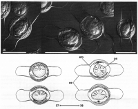
Eggs of Proteocephalus synodontis Woodland, 1925 from Synodontis schall, Lake Turkana, Kenya (36, 37) and S. schall, Khartoum, Sudan (38). Abbreviations: em - embryophore; oe - outer envelope; on-onco... MoreEggs of Proteocephalus synodontis Woodland, 1925 from Synodontis schall, Lake Turkana, Kenya (36, 37) and S. schall, Khartoum, Sudan (38). Abbreviations: em - embryophore; oe - outer envelope; on-oncosphere. Scale bars = 20 μm. |
Scanning Electron Micrograph
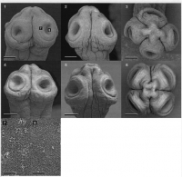
FIGURES 1-13. Scanning electron micrographs of the scoleces of Proteocephalus spp. from Synodontis catfish: P synodontis
Woodland, 1925 from S. schall, Lake Turkana, Kenya (1-3, 7, 8) and from S. sch... MoreFIGURES 1-13. Scanning electron micrographs of the scoleces of Proteocephalus spp. from Synodontis catfish: P synodontis
Woodland, 1925 from S. schall, Lake Turkana, Kenya (1-3, 7, 8) and from S. schall, Kosti, Sudan (4-6); P membranacei
Troncy, 1978 from S. membranacea, Lake Chad, Chad, syntype (MNHNP 1116H; 9, 10); P largoproglottis (= syn. ofP membranacei),
syntype (MNHNP 1115H; 11-13). 1,4,9,11. Dorsoventral view. 2, 5,10,13. Lateral view. 3, 6,12. Apical view. 7,
8. Papilliform filitriches on the external rim and internal surface of suckers, respectively (see Fig. 1). Scale bars = 100 µm (1-6,
9,11-13); 50 µm (10); 3 µm (7,8). |
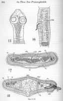
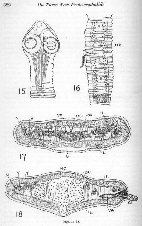 Figs. 15-18. Proteocephalus synodontis.
G, cortex; GI, cirrus; DU, diverticula of uterus; IL, internal layer (sheath) of longitudina
muscles; MG, median chamber of uterus; N, nerve; OV, ovary; T, testes; UD, uterine
canal; UTO, opening of uterus to exterior; V, vitellaria; VA, vagina.
Fig. 15. Scolex in outline. (x 52.)
Fig. 16. Semi.transparent view of an elongated ripe proglottid. (x 36.)
Fig. 17. Transverse section across a ripe proglottid in the region of the ovary. Note in this and
the next figure the enormous development of the internal longitudinal muscle sheath, which
obliterates the cortex save for a small area in the median ventral line. (x 74.)
Fig. 18. Transverse section across a ripe proglottid in the region of the cirrus sac. (x74.)
Figs. 15-18. Proteocephalus synodontis.
G, cortex; GI, cirrus; DU, diverticula of uterus; IL, internal layer (sheath) of longitudina
muscles; MG, median chamber of uterus; N, nerve; OV, ovary; T, testes; UD, uterine
canal; UTO, opening of uterus to exterior; V, vitellaria; VA, vagina.
Fig. 15. Scolex in outline. (x 52.)
Fig. 16. Semi.transparent view of an elongated ripe proglottid. (x 36.)
Fig. 17. Transverse section across a ripe proglottid in the region of the ovary. Note in this and
the next figure the enormous development of the internal longitudinal muscle sheath, which
obliterates the cortex save for a small area in the median ventral line. (x 74.)
Fig. 18. Transverse section across a ripe proglottid in the region of the cirrus sac. (x74.) 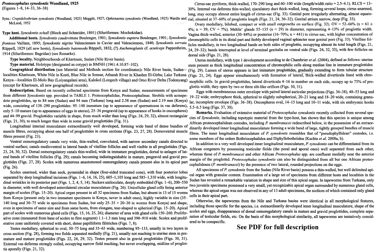
 FIGURES 14-24. Proteocephalus synodontis Woodland, 1925. 14, Holotype (BMNH 1961.4.10.87-102), Khartoum, Sudan,
'&:olex, dorsoventral view. IS, 17-20. Apical organs. IS. Holotype, Khartoum. 17. From Synodontis schall, Girba, Sudan; 18.
Immature tapeworm from Synodontis schall, Lake Turkana, Kenya; 19. From Synodontis schall, Kostl, Sudan; 20. From Synodontis
caudovittata, Kostl, Sudan. 21, 24. Immature proglottides from S. caudovittata, Kosti, Sudan. 22. Holotype, immature
proglottis (note median extent oftestes not reaching to uterine stem). 23. Holotype, cross section of gravid proglottis. Abbreviations:
:ur-apical organ; cs---eirrus-sac; do--dorsal osmoregulatory canals; dv-dorsoventral muscle fibres; gc-gland cells;
Im-loogitudinal internal musculature; ov-ovary; sd-sperm duct (vas deferens); te-testes; ud-uterine diverticulum; uourerine
orifice; ut-oterus; vc-vaginal canal; vi-vitelline follicles; vo-ventral osmoregulatory canals. Scale bars = 100 µm
(I ,16, _1--' 4); 50 µm (IS, 17-20).
FIGURES 14-24. Proteocephalus synodontis Woodland, 1925. 14, Holotype (BMNH 1961.4.10.87-102), Khartoum, Sudan,
'&:olex, dorsoventral view. IS, 17-20. Apical organs. IS. Holotype, Khartoum. 17. From Synodontis schall, Girba, Sudan; 18.
Immature tapeworm from Synodontis schall, Lake Turkana, Kenya; 19. From Synodontis schall, Kostl, Sudan; 20. From Synodontis
caudovittata, Kostl, Sudan. 21, 24. Immature proglottides from S. caudovittata, Kosti, Sudan. 22. Holotype, immature
proglottis (note median extent oftestes not reaching to uterine stem). 23. Holotype, cross section of gravid proglottis. Abbreviations:
:ur-apical organ; cs---eirrus-sac; do--dorsal osmoregulatory canals; dv-dorsoventral muscle fibres; gc-gland cells;
Im-loogitudinal internal musculature; ov-ovary; sd-sperm duct (vas deferens); te-testes; ud-uterine diverticulum; uourerine
orifice; ut-oterus; vc-vaginal canal; vi-vitelline follicles; vo-ventral osmoregulatory canals. Scale bars = 100 µm
(I ,16, _1--' 4); 50 µm (IS, 17-20). 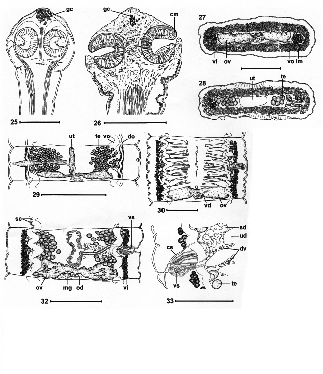 FIGURES 25-35. Proteocephalus synodontis Woodland, 1925 from Synodontis schall, Lake Turkana, Kenya (25-33) and P
membranacei from S. membranacea, Lake Chad, Chad. 25. Scolex, dorsoventral view (note accumulation of gland cells in the
apical region-gc). 26. Scolex, longitudinal section. 27, 28, 34, 35. Cross sections of pregravid proglottides at the level of
ovary (27, 34), cirrus-sac (28) and testes (35). 29. Premature proglottis, dorsal view. 30, 31. Gravid proglottides of different
shape, ventral view. 32. Pregravid proglottis, ventral view. 33. Terminal genitalia, ventral view (note vaginal sphincter-vs).
Fig. 34-syntype of P membranacei (MNHNP 1116H); Fig. 35-syntype of P largoproglottis (= syn. of P membranacei;
MNHNP 1115H). Abbreviations: cm---eircular musculature of suckers; cs---eirrus-sac; do--dorsal osmoregulatory canals;
dv---dorsoventral muscle fibres; gc-gland cells; 1m-longitudinal internal musculature; mg-Mehlis' gland; od-{)viduct;
ov-{)vary; sc-secondary osmoregulatory canals; sd-sperm duct (vas deferens); te--testes; ud-uterine diverticulum; uouterine
orifice; ut-uterus; vd-vitelloduct; vi-vitelline follicles; vo-ventral osmoregulatory canals; vs-vaginal sphincter.
Scale bars = 500 µm (25-32, 34, 35); 250 µm (33).
FIGURES 25-35. Proteocephalus synodontis Woodland, 1925 from Synodontis schall, Lake Turkana, Kenya (25-33) and P
membranacei from S. membranacea, Lake Chad, Chad. 25. Scolex, dorsoventral view (note accumulation of gland cells in the
apical region-gc). 26. Scolex, longitudinal section. 27, 28, 34, 35. Cross sections of pregravid proglottides at the level of
ovary (27, 34), cirrus-sac (28) and testes (35). 29. Premature proglottis, dorsal view. 30, 31. Gravid proglottides of different
shape, ventral view. 32. Pregravid proglottis, ventral view. 33. Terminal genitalia, ventral view (note vaginal sphincter-vs).
Fig. 34-syntype of P membranacei (MNHNP 1116H); Fig. 35-syntype of P largoproglottis (= syn. of P membranacei;
MNHNP 1115H). Abbreviations: cm---eircular musculature of suckers; cs---eirrus-sac; do--dorsal osmoregulatory canals;
dv---dorsoventral muscle fibres; gc-gland cells; 1m-longitudinal internal musculature; mg-Mehlis' gland; od-{)viduct;
ov-{)vary; sc-secondary osmoregulatory canals; sd-sperm duct (vas deferens); te--testes; ud-uterine diverticulum; uouterine
orifice; ut-uterus; vd-vitelloduct; vi-vitelline follicles; vo-ventral osmoregulatory canals; vs-vaginal sphincter.
Scale bars = 500 µm (25-32, 34, 35); 250 µm (33). 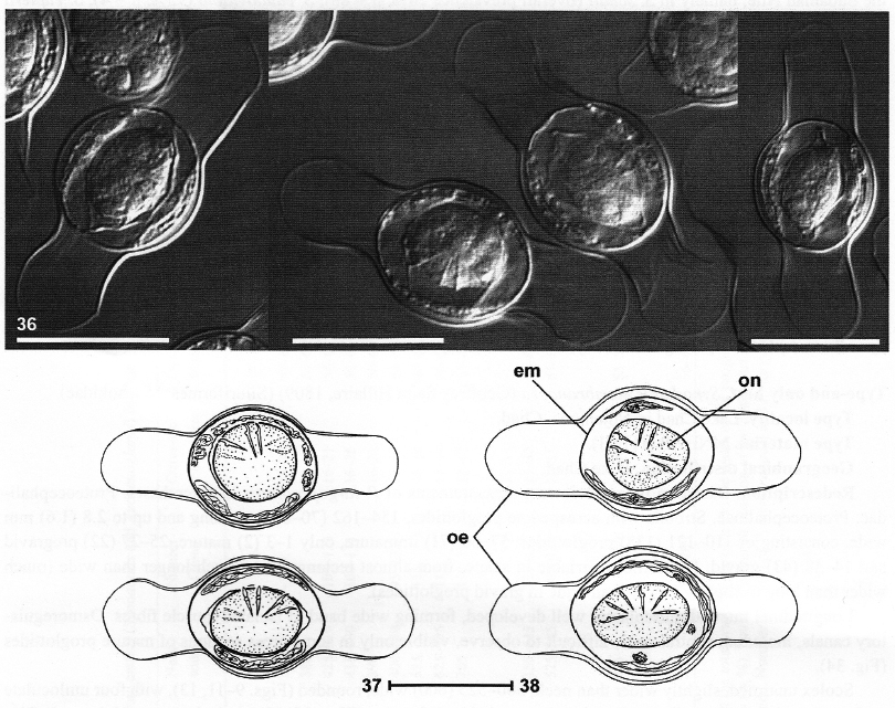 Eggs of Proteocephalus synodontis Woodland, 1925 from Synodontis schall, Lake Turkana, Kenya (36, 37) and S. schall, Khartoum, Sudan (38). Abbreviations: em - embryophore; oe - outer envelope; on-oncosphere. Scale bars = 20 μm.
  FIGURES 1-13. Scanning electron micrographs of the scoleces of Proteocephalus spp. from Synodontis catfish: P synodontis
Woodland, 1925 from S. schall, Lake Turkana, Kenya (1-3, 7, 8) and from S. schall, Kosti, Sudan (4-6); P membranacei
Troncy, 1978 from S. membranacea, Lake Chad, Chad, syntype (MNHNP 1116H; 9, 10); P largoproglottis (= syn. ofP membranacei),
syntype (MNHNP 1115H; 11-13). 1,4,9,11. Dorsoventral view. 2, 5,10,13. Lateral view. 3, 6,12. Apical view. 7,
8. Papilliform filitriches on the external rim and internal surface of suckers, respectively (see Fig. 1). Scale bars = 100 µm (1-6,
9,11-13); 50 µm (10); 3 µm (7,8).
FIGURES 1-13. Scanning electron micrographs of the scoleces of Proteocephalus spp. from Synodontis catfish: P synodontis
Woodland, 1925 from S. schall, Lake Turkana, Kenya (1-3, 7, 8) and from S. schall, Kosti, Sudan (4-6); P membranacei
Troncy, 1978 from S. membranacea, Lake Chad, Chad, syntype (MNHNP 1116H; 9, 10); P largoproglottis (= syn. ofP membranacei),
syntype (MNHNP 1115H; 11-13). 1,4,9,11. Dorsoventral view. 2, 5,10,13. Lateral view. 3, 6,12. Apical view. 7,
8. Papilliform filitriches on the external rim and internal surface of suckers, respectively (see Fig. 1). Scale bars = 100 µm (1-6,
9,11-13); 50 µm (10); 3 µm (7,8). 

