Cestode Scientific Name
| Species ID | 12557 |
|---|---|
| Order | Onchoproteocephalidea I |
| Family | Proteocephalidae |
| Subfamily | Proteocephalinae |
| Genus | Solenotaenia |
| Species | viperis |
| Authority | Beddard, 1913 |
| Taxonomic Status | Synonym |
| Valid Name | Crepidobothrium viperis (Beddard, 1913) Meggitt, 1927 |
| Synonyms | Ophiotaenia viperis (Beddard, 1913) Rudin, 1917; Proteocephalus viperis (Beddard, 1913) Woodland, 1925 |
| Genus Record | No |
| Type Species | |
| Verified | Yes |
| Verified By | T. Scholz |
| Citation(s) |
Beddard, F. E. 1913. Contributions to the anatomy and systematic arrangement of the Cestoidea. IX. On a new genus of Ichthytaeniids. Proceedings of the Zoological Society of London 1913(1): 243-261. (3971) Download PDF |
| Redescription | |
| Scientific Name Notes |
Record Data
| Date (MM/DD/YYYY) | Action | User Name |
|---|---|---|
| 02/14/2013 | Created | B. Barbeau |
| 10/08/2013 | Modified | |
| 05/18/2020 | Modified | T. Scholz |
| 12/03/2021 | Modified | B. Barbeau |
Type Host
| Host Class | Reptilia | ||||||
|---|---|---|---|---|---|---|---|
| Host Order | Squamata | ||||||
| Host Family | Viperidae | ||||||
|
Type Host (Literal) |
|
||||||
|
Type Host (Valid) |
|
||||||
| Additional Host(s) | |||||||
| Site in Host | |||||||
| Host Notes |
Type Locality
| Country | South America |
|---|---|
| Body of Water | |
| Island(s) | |
| City/Region | |
| Coordinates | |
| DD Latitude | |
| DD Longitude | |
| Additional Localities | |
| Locality Notes |
Specimens
| Type Material | |
|---|---|
| Total Number of Type Specimens | |
| Voucher Material | |
| Specimen Notes |
Data are given as in original description unless otherwise indicated.
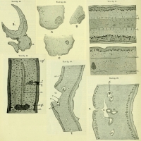
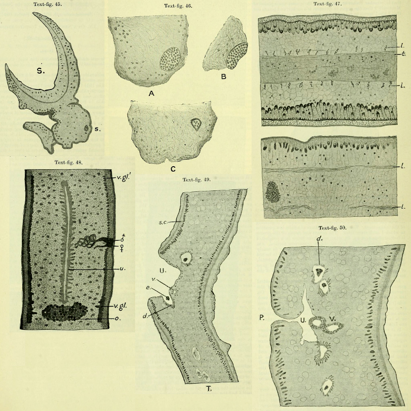 Text-fig. 45. Transverse section through scolex of Solenotaenia viperis. S. One of four large suckers. s. Small apical sucker showing its minute size in comparison with large suckers. Text-fig. 46. Three nearly consecutive sections through minute apical sucker of Solenotaenia viperis. A. At a little distance from the surface. B. At the actual apex of the scolex. C. Near to deep end of apical sucker. Text-fig. 47. Upper figure a transverse section through part of a proglottid of Solenotaenia viperis; lower figure a longitudinal section through part of a proglottid. l. Longitudinal muscles. t. Transverse muscles. Text-fig. 48. View of an entire proglottid of Solenotaenia viperis stained and mounted as a transparent object. ♂, opening of cirrus-sac; ♀, opening of vagina. Text-fig. 49. Transverse section through middle region of proglottid of Solenotaenia viperis, to show the ventral uterine groove. d. Diverticulum of uterus containing an embryo (e). The opening into the uterus of the corresponding diverticulum is not shown on the opposite side. sc. Subcuticular layer. T. Testis. U. Uterus. v. Vagina. Text-fig. 50. Transverse section through middle of a proglottid of Solenotaenia viperis at posterior end of uterine groove. d. Diverticula of uterus containing embryos. P. External pore which forms the commencement of the ventral uterine groove. U. Uterus. V. Vagina, which is seen cut through twice, being coiled in this region near to the ovary.
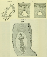
 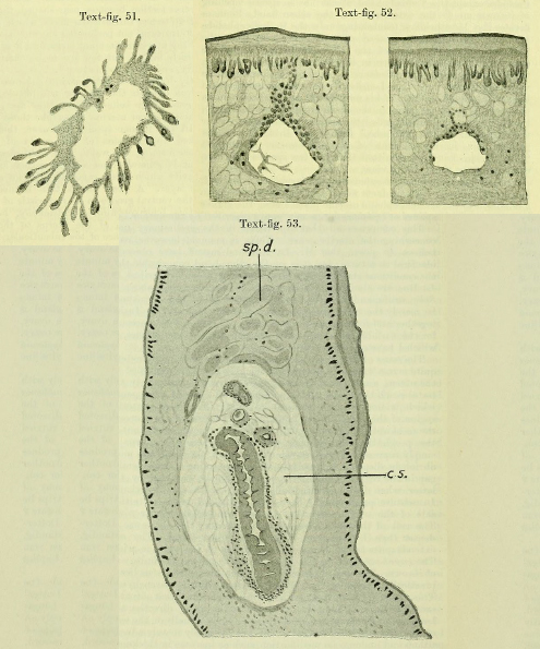 Text-fig. 51. A transverse section of one of the uterine diverticula of Solenotaenia viperis highly magnified to show the long-stalked glandular cells which form the greater part of its walls. Text-fig. 52. Two nearly consecutive sections through unripe uterus of Solenotaenia viperis showing (in the left-hand figure) a rudimentary external pore. The lateral, thickenings of the uterine tube are probably the commencement of the lateral diverticula. Text-fig. 53. Transverse section through a portion of a proglottid of Solenotaenia viperis, showing a cirrus-sac, c.s., which is seen to have no definite walls. In the interior of the sac is shown the terminal wider part of the cirrus and three sections of the narrower part of that tube. sp.d. Coiled region of sperm-duct.
 Best viewed in Firefox


