Cestode Scientific Name
| Species ID | 12505 |
|---|---|
| Order | Onchoproteocephalidea I |
| Family | |
| Subfamily | |
| Genus | Ophiotaenia |
| Species | gallardi |
| Authority | (Johnston 1911) Freze, 1965 |
| Taxonomic Status | Valid |
| Valid Name | |
| Synonyms | |
| Genus Record | No |
| Type Species | |
| Verified | No |
| Verified By | |
| Citation(s) |
Johnston, T. H. 1911. Proteocephaus gallardi. A new cestode from the black snake. Annals of the Queensland Museum 10: 175-182. (4099) Download PDFFreze, V. I. 1965. [Proteocephalata in Fish, Amphibians, and Reptiles] (In Russian). Osnovy Tsestodologii 5: 538 pp. (4031) Download PDF |
| Redescription |
de Chambrier, S. and A. de Chambrier. 2010. Two new genera and two new species of proteocephalidean tapeworms (Eucestoda) from reptiles and amphibians in Australia. Folia Parasitologica 57(4): 263-279. (6026) Download PDF |
| Scientific Name Notes |
Record Data
| Date (MM/DD/YYYY) | Action | User Name |
|---|---|---|
| 02/14/2013 | Created | B. Barbeau |
| 06/10/2014 | Modified | |
| 01/30/2019 | Modified | R. Kuchta |
| 06/24/2020 | Modified | B. Barbeau |
Type Host
| Host Class | |||||||
|---|---|---|---|---|---|---|---|
| Host Order | |||||||
| Host Family | |||||||
|
Type Host (Literal) |
|
||||||
|
Type Host (Valid) |
|
||||||
| Additional Host(s) | |||||||
| Site in Host | intestine | ||||||
| Host Notes |
Type Locality
| Country | Australia |
|---|---|
| Body of Water | |
| Island(s) | |
| City/Region | |
| Coordinates | |
| DD Latitude | |
| DD Longitude | |
| Additional Localities | |
| Locality Notes | New South Wales and Victoria |
Specimens
| Type Material | Lectotype QM G12/110, Gippsland, Victoria, Australia, A.S. Le Seouf 1910. Paralextotypes which are conspecific with the lectotype: QM G12/108-109, Narrara Near Gosford, NSW, Australia, L. Gallard 1910; AHC 20081 (= SAM 2295 in part), Victoria, collected by A.S. Le Souef. |
|---|---|
| Total Number of Type Specimens | Based on 7 specimens |
| Voucher Material | |
| Specimen Notes | Lectotype QM G12/110, Gippsland, Victoria, Australia, A.S. Le Seouf 1910. Paralectotypes which are conspecific with the lectotype: QM G12/108-109, Narrara Near Gosford, NSW, Australia, L. Gallard 1910; AHC 20081 (= SAM 2295 in part), Victoria, collected by A.S. Le Souef. As per the redescription by de Chambreir, S. and A. de Chambreir. |
Data are given as in original description unless otherwise indicated.
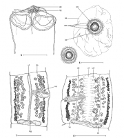

 Figs. 58. Ophiotaenia gallardi (Johnston, 1911). Fig. 5. Syntype, scolex, ventral view (QM G12/110) (redrawn from Johnston
1911). Fig. 6. Eggs in distilled water, showing the three-layered
embryophore and small outgrowths (SAM 4374). Fig. 7. Syntype, mature proglottis, dorsal view (QM G12/109) (redrawn from Johnston 1911) (ventral osmoregulatory canal not figured).
Fig. 8. Pregravid proglottis, ventral view (SAM 4374 b); note the position of dorsal osmoregulatory canals, which separate longitudinally the testicular fields. Abbreviations: ao - apical organ; cg - cells with finely granular cytoplasm; do - dorsal osmoregulatory canal; em three-layered embryophore; mg Mehlis glands; oe outer envelope; on oncosphere; ov ovary: so small outgrowths; ud - uterine diverticula; ut - uterus; va - vas deferens; vo - ventral osmoregulatory canal. Scale bar: Figs. 5, 7 = 500µm; Fig. 6 = 50µm; Fig. 8 = 1mm.
Figs. 58. Ophiotaenia gallardi (Johnston, 1911). Fig. 5. Syntype, scolex, ventral view (QM G12/110) (redrawn from Johnston
1911). Fig. 6. Eggs in distilled water, showing the three-layered
embryophore and small outgrowths (SAM 4374). Fig. 7. Syntype, mature proglottis, dorsal view (QM G12/109) (redrawn from Johnston 1911) (ventral osmoregulatory canal not figured).
Fig. 8. Pregravid proglottis, ventral view (SAM 4374 b); note the position of dorsal osmoregulatory canals, which separate longitudinally the testicular fields. Abbreviations: ao - apical organ; cg - cells with finely granular cytoplasm; do - dorsal osmoregulatory canal; em three-layered embryophore; mg Mehlis glands; oe outer envelope; on oncosphere; ov ovary: so small outgrowths; ud - uterine diverticula; ut - uterus; va - vas deferens; vo - ventral osmoregulatory canal. Scale bar: Figs. 5, 7 = 500µm; Fig. 6 = 50µm; Fig. 8 = 1mm. 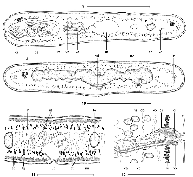 Figs. 912. Ophiotaenia gallardi (Johnston, 1911). Figs. 9, 10. Syntype (SAM 437416,
9). Cross-sections
at the level of the cirrus-sac and ovary, respectively. Fig. 11. Crosssection
at the level of the anterior part of pregravid proglottis (SAM 4375). Fig. 12. Vagina and cirrussac
region, ventral view (SAM 4374c). Abbreviations: ci cirrus; cs cirrussac;
do dorsal osmoregulatory canal; lm internal longitudinal musculature; ln longitudinal nerve; mi microtriches; ov ovary; sc subtegumental cells; st subtegumental muscle fibres; te - testes; tg - tegument; up - uterine pore-like; ut - uterus; va - vas deferens; vc - vaginal canal; vd - vitelloduct; vi - vitelline follicles; vo - ventral osmoregulatory canal; vs - vaginal sphincter. Scale bars: Figs. 9, 10, 12 = 500µm; Fig. 11 = 250µm.
Figs. 912. Ophiotaenia gallardi (Johnston, 1911). Figs. 9, 10. Syntype (SAM 437416,
9). Cross-sections
at the level of the cirrus-sac and ovary, respectively. Fig. 11. Crosssection
at the level of the anterior part of pregravid proglottis (SAM 4375). Fig. 12. Vagina and cirrussac
region, ventral view (SAM 4374c). Abbreviations: ci cirrus; cs cirrussac;
do dorsal osmoregulatory canal; lm internal longitudinal musculature; ln longitudinal nerve; mi microtriches; ov ovary; sc subtegumental cells; st subtegumental muscle fibres; te - testes; tg - tegument; up - uterine pore-like; ut - uterus; va - vas deferens; vc - vaginal canal; vd - vitelloduct; vi - vitelline follicles; vo - ventral osmoregulatory canal; vs - vaginal sphincter. Scale bars: Figs. 9, 10, 12 = 500µm; Fig. 11 = 250µm.  Figs.
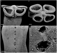
14. Ophiotaenia gallardi (Johnston, 1911). Scanning electron micrographs. Fig. 1. Scolex, dorsoventral view (MHNG INVE
67494). Fig. 2. Scolex, apical view (MHNG INVE 67494). Fig. 3. Ventral view of a gravid proglottis, showing uterine pore-like
structures (SAM 4375). Fig. 4. Detail of uterine pore-like structures (SAM 4375). Scale bars: Figs. 1, 2 = 100µm; Fig. 3 = 200 µm; Fig. 4 = 20 µm.
Figs.
14. Ophiotaenia gallardi (Johnston, 1911). Scanning electron micrographs. Fig. 1. Scolex, dorsoventral view (MHNG INVE
67494). Fig. 2. Scolex, apical view (MHNG INVE 67494). Fig. 3. Ventral view of a gravid proglottis, showing uterine pore-like
structures (SAM 4375). Fig. 4. Detail of uterine pore-like structures (SAM 4375). Scale bars: Figs. 1, 2 = 100µm; Fig. 3 = 200 µm; Fig. 4 = 20 µm. Best viewed in Firefox


