Cestode Scientific Name
| Species ID | 12502 |
|---|---|
| Order | Onchoproteocephalidea I |
| Family | Proteocephalidae |
| Subfamily | Acanthotaeniinae |
| Genus | Proteocephalus |
| Species | beddardi |
| Authority | Woodland, 1925 |
| Taxonomic Status | Synonym |
| Valid Name | Acanthotaenia beddardi (Woodland, 1925) Schmidt, 1986 |
| Synonyms | Crepidobothrium beddardi (Woodland, 1925) Meggitt, 1927; Rostellotaenia beddardi (Woodland, 1925) Freze, 1963 |
| Genus Record | No |
| Type Species | No |
| Verified | Yes |
| Verified By | T. Scholz |
| Citation(s) |
Woodland, W. N. F. 1925. On three new proteocephalids (Cestoda) and a revision of the genera of the family. Parasitology 17: 370-394. (3958) Download PDF |
| Redescription | |
| Scientific Name Notes |
Record Data
| Date (MM/DD/YYYY) | Action | User Name |
|---|---|---|
| 02/14/2013 | Created | B. Barbeau |
| 11/08/2013 | Modified | |
| 02/04/2020 | Modified | B. Barbeau |
| 03/09/2020 | Modified | P. Lopez Dineen |
| 04/07/2020 | Modified | T. Scholz |
| 12/03/2021 | Modified | B. Barbeau |
Type Host
| Host Class | |||||||
|---|---|---|---|---|---|---|---|
| Host Order | |||||||
| Host Family | |||||||
|
Type Host (Literal) |
|
||||||
|
Type Host (Valid) |
|
||||||
| Additional Host(s) | |||||||
| Site in Host | |||||||
| Host Notes |
Type Locality
| Country | |
|---|---|
| Body of Water | |
| Island(s) | |
| City/Region | |
| Coordinates | |
| DD Latitude | |
| DD Longitude | |
| Additional Localities | |
| Locality Notes |
Specimens
| Type Material | |
|---|---|
| Total Number of Type Specimens | |
| Voucher Material | |
| Specimen Notes |
Data are given as in original description unless otherwise indicated.

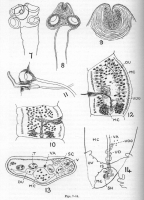
 Figs. 7-14. Proteocephalus beddardi. Fig. 7. Scolex in outline. (x 116). Fig. 8. Another view of the scolex showing the apical organ (muscular rostellum?). (x 116). Fig. 9. The apical organ seen in optical section, with the spinelets. (x 346). Fig. 10. A young mature proglottid. Dorsal view. (x 52.) Fig. 11. The cirrus sac, armed cirrus and vaginal opening from the ventral aspect. The vaginal opening is somewhat distorted. (x 74.) Fig. 12. Ripe proglottid, much flattened, in dorsal view. Note the wide median chamber of the uterus sac and the presence of the uterus below the ovary. (x 23). Fig. 13. Transverse section (anterior to the uterine canal) across a young ripe proglottid. Note absence of the internal longitudinal muscle sheath. (x 116). Fig. 14. The ducts in the region of the ovarian isthmus, viewed from the dorsal side. (x 74.) DU, diverticula of uterus; IS, isthmus of ovary; MC, median chamber of uterus; OV; ovary; SC, nuclear layer of subcuticula; SH, shell gland; T, testes; UD, uterine canal; UDO, opening of uterine canal into median chamber of uterus sac; V, vitellaria; VA, vagina.
 Best viewed in Firefox


