Line Drawing 1

FIG. 1. Diphyllobothrium ursi n. sp., mature segment from naturally infected brown bear, Kodiak Island, Alaska.
FIG. 2. D. ursi n. sp., mature segment from naturally infected brown bear.
FIG. 3. D. ... MoreFIG. 1. Diphyllobothrium ursi n. sp., mature segment from naturally infected brown bear, Kodiak Island, Alaska.
FIG. 2. D. ursi n. sp., mature segment from naturally infected brown bear.
FIG. 3. D. ursi n. sp., scolex of mature cestode from naturally infected brown bear.
FIG. 4. D. ursi n. sp., mature segment from naturally infected brown bear.
FIG. 5. D. ursi n. sp., sagittal section through mature segment, showing relationships of genital ducts. From naturally infected brown bear.
FIG. 6. D. ursi n. sp., plerocercoid larva from red salmon, excysted.
FIG. 7. D. ursi n. sp., 10-day-old mature segment from experimentally infected black bear.
FIG. 8. D. ursi n. sp., 61-day-old mature segment from experimentally infected black bear. |
Line Drawing 2

FIG. 10. D. ursi n. sp., scolex of plerocercoid, lateral view.
FIG. 11. D. ursi n. sp., segment of cross-section of plerocercoid; drawn from 5 micron section, stained with hematoxylin-eosin.
FIG. 12... MoreFIG. 10. D. ursi n. sp., scolex of plerocercoid, lateral view.
FIG. 11. D. ursi n. sp., segment of cross-section of plerocercoid; drawn from 5 micron section, stained with hematoxylin-eosin.
FIG. 12. D. ursi n. sp., scolex of plerocercoid, dorso-ventral view. |
Photo Micrograph
|
Scanning Electron Micrograph
|
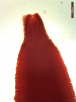

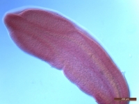
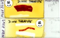
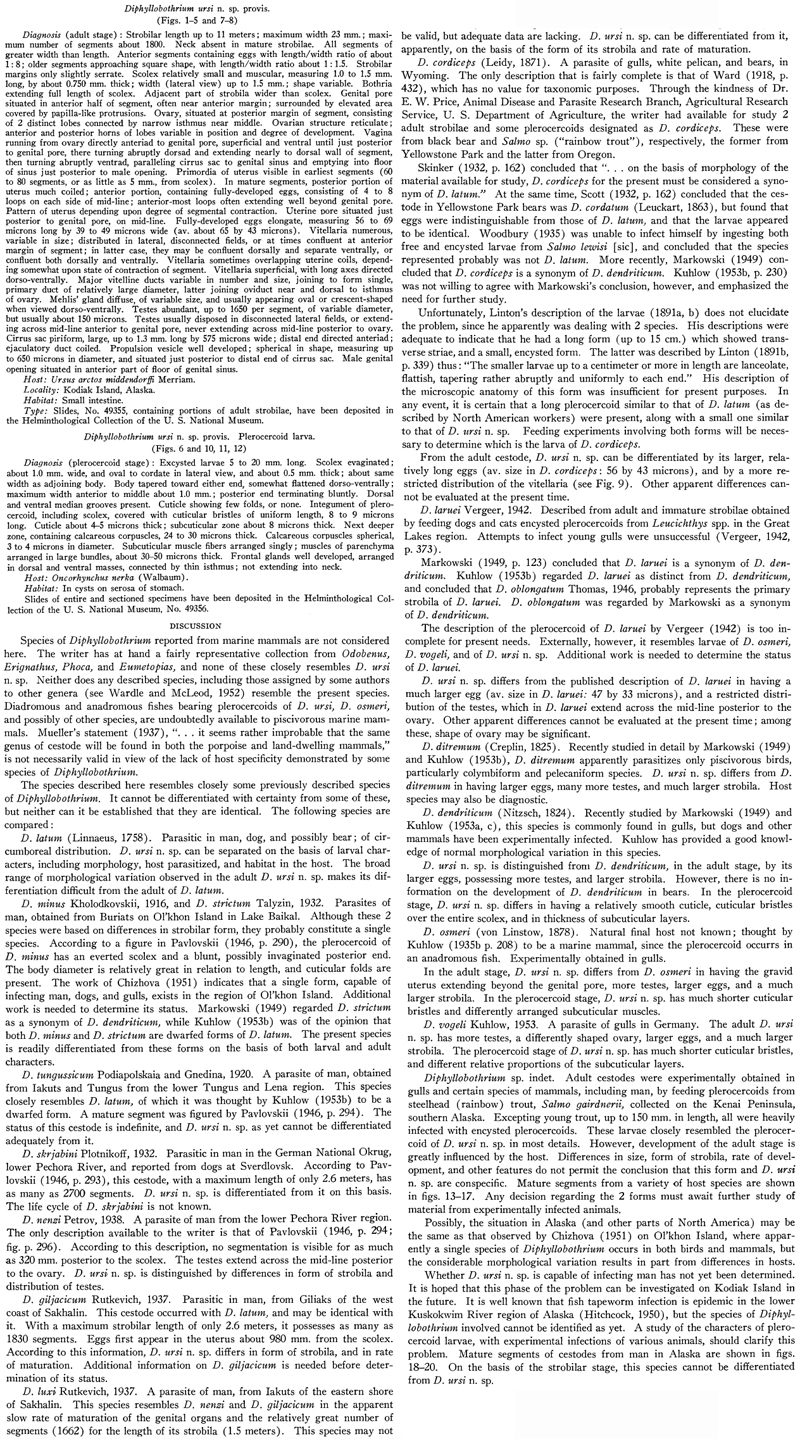
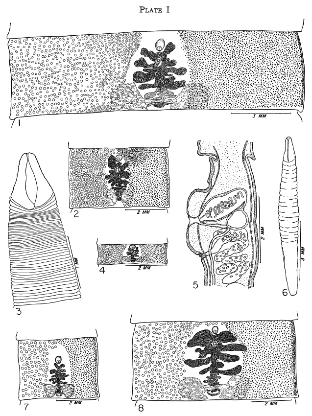 FIG. 1. Diphyllobothrium ursi n. sp., mature segment from naturally infected brown bear, Kodiak Island, Alaska.
FIG. 2. D. ursi n. sp., mature segment from naturally infected brown bear.
FIG. 3. D. ursi n. sp., scolex of mature cestode from naturally infected brown bear.
FIG. 4. D. ursi n. sp., mature segment from naturally infected brown bear.
FIG. 5. D. ursi n. sp., sagittal section through mature segment, showing relationships of genital ducts. From naturally infected brown bear.
FIG. 6. D. ursi n. sp., plerocercoid larva from red salmon, excysted.
FIG. 7. D. ursi n. sp., 10-day-old mature segment from experimentally infected black bear.
FIG. 8. D. ursi n. sp., 61-day-old mature segment from experimentally infected black bear.
FIG. 1. Diphyllobothrium ursi n. sp., mature segment from naturally infected brown bear, Kodiak Island, Alaska.
FIG. 2. D. ursi n. sp., mature segment from naturally infected brown bear.
FIG. 3. D. ursi n. sp., scolex of mature cestode from naturally infected brown bear.
FIG. 4. D. ursi n. sp., mature segment from naturally infected brown bear.
FIG. 5. D. ursi n. sp., sagittal section through mature segment, showing relationships of genital ducts. From naturally infected brown bear.
FIG. 6. D. ursi n. sp., plerocercoid larva from red salmon, excysted.
FIG. 7. D. ursi n. sp., 10-day-old mature segment from experimentally infected black bear.
FIG. 8. D. ursi n. sp., 61-day-old mature segment from experimentally infected black bear. 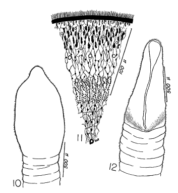 FIG. 10. D. ursi n. sp., scolex of plerocercoid, lateral view.
FIG. 11. D. ursi n. sp., segment of cross-section of plerocercoid; drawn from 5 micron section, stained with hematoxylin-eosin.
FIG. 12. D. ursi n. sp., scolex of plerocercoid, dorso-ventral view.
FIG. 10. D. ursi n. sp., scolex of plerocercoid, lateral view.
FIG. 11. D. ursi n. sp., segment of cross-section of plerocercoid; drawn from 5 micron section, stained with hematoxylin-eosin.
FIG. 12. D. ursi n. sp., scolex of plerocercoid, dorso-ventral view.  -USNPC No. 49355
 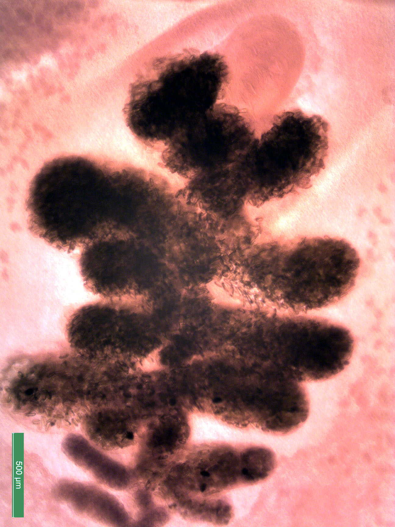 -USNPC No. 49355
  -USNPC No. 49356 (plerocercoid)
  MSBP No. 5733

