Line Drawing 1

Fig. 33. Young segments.
Fig. 34. Scolex lateral view.
Fig. 35. Segments, with genitalia.
Fig. 36. Genitalia.
Fig. 37. Transverse section of segment.
a.s., accessory saccnlus; b., bothrium; c.,... MoreFig. 33. Young segments.
Fig. 34. Scolex lateral view.
Fig. 35. Segments, with genitalia.
Fig. 36. Genitalia.
Fig. 37. Transverse section of segment.
a.s., accessory saccnlus; b., bothrium; c., cirrus; c.s., cirrus-sac; c.b., cavity of bothrium; e.g.; cement glands; d.e.v., dorsal excretory vessels;; d.v.m., dorso-ventral muscle; e., egg; e.c., egg capsule; e.p., excretory pore; e.v., excretory vessel ; g.c., genital cloaca; g.p., genital pore; h., hook; l., lemniscus; l.m., l.m.l., l.m. 2, longitudinal muscles; m.b., male bursa; n., longitudinal nerve; ov., ovary; p.s., proboscis sheath; r., rostellum; r.c., rostellar sac; r.s., receptaculum seminis; s., sphincter muscle surrounding genital cloaca; s.g., shell gland; s.l., suspensory ligament; t., testis; t.e.v., transverse excretory vessel; tr. m., transverse muscle; u., uterus; u.b., uterine bell; u.a., uterine aperture; u.d., uterine duct; ·v., vagina; v.d., vas deferens; v.e.v., Yentral excretory vessels; v.g., vitelline gland; v.s., vesicnla seminalis; vt. d., vitelline duct. |
Line Drawing 2
|
Photo Micrograph
|
Scanning Electron Micrograph
|
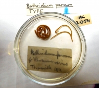
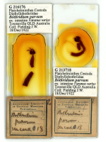
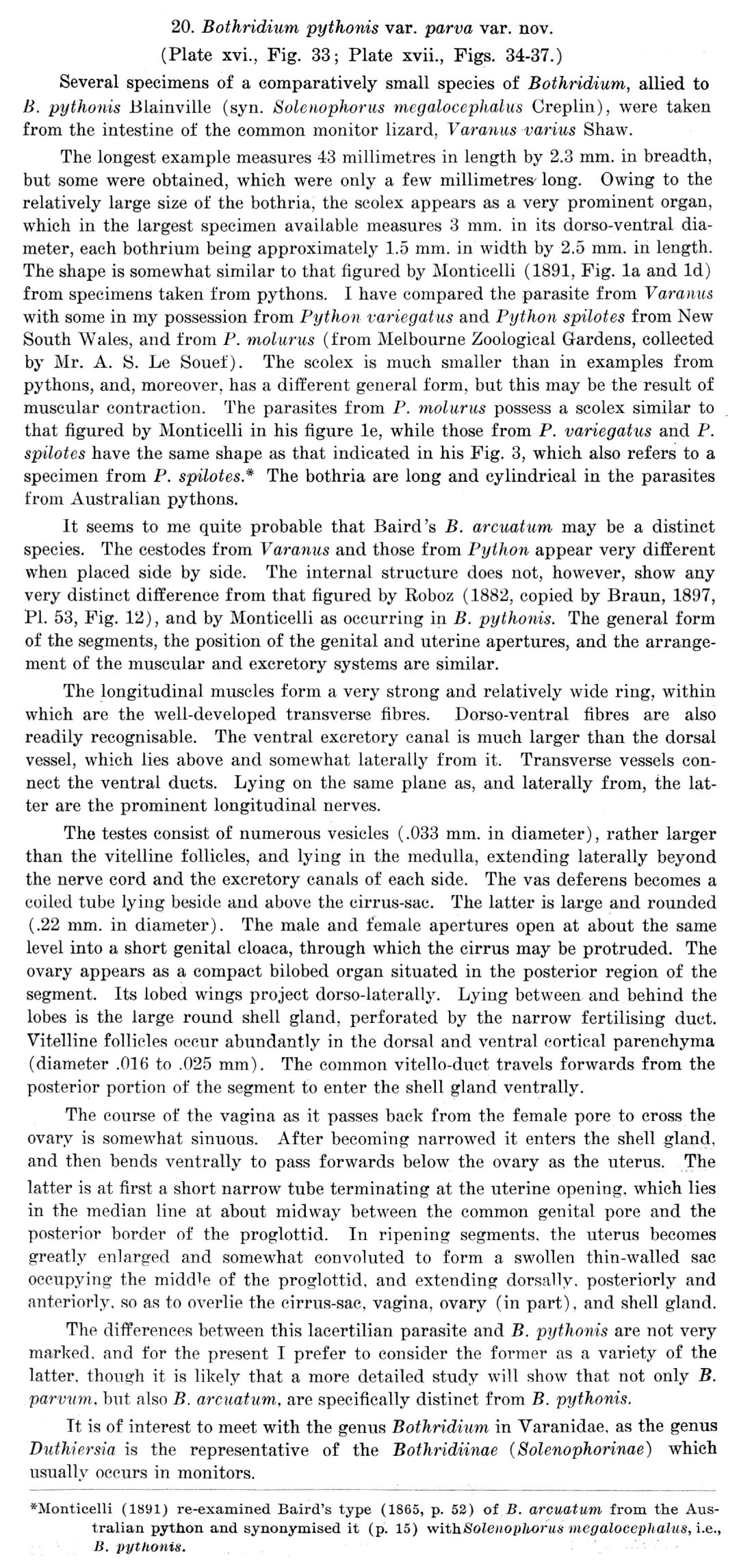
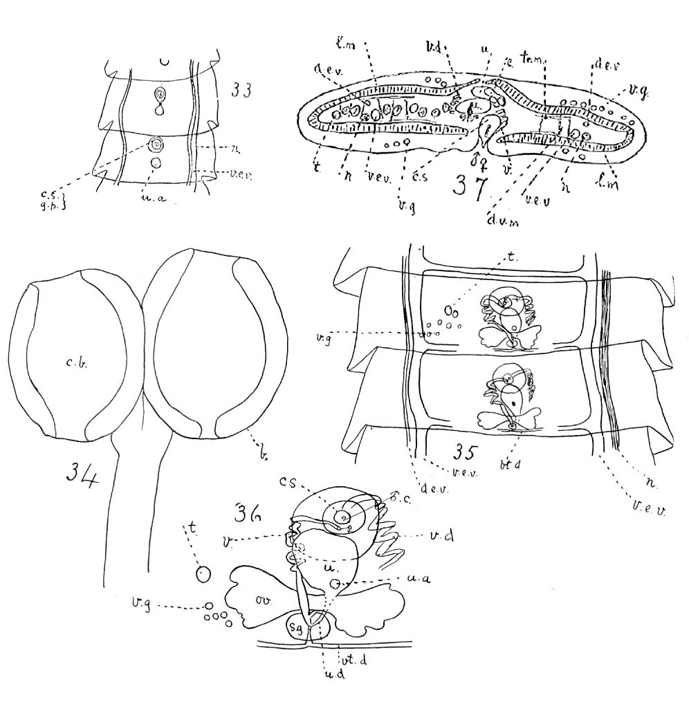 Fig. 33. Young segments.
Fig. 34. Scolex lateral view.
Fig. 35. Segments, with genitalia.
Fig. 36. Genitalia.
Fig. 37. Transverse section of segment.
a.s., accessory saccnlus; b., bothrium; c., cirrus; c.s., cirrus-sac; c.b., cavity of bothrium; e.g.; cement glands; d.e.v., dorsal excretory vessels;; d.v.m., dorso-ventral muscle; e., egg; e.c., egg capsule; e.p., excretory pore; e.v., excretory vessel ; g.c., genital cloaca; g.p., genital pore; h., hook; l., lemniscus; l.m., l.m.l., l.m. 2, longitudinal muscles; m.b., male bursa; n., longitudinal nerve; ov., ovary; p.s., proboscis sheath; r., rostellum; r.c., rostellar sac; r.s., receptaculum seminis; s., sphincter muscle surrounding genital cloaca; s.g., shell gland; s.l., suspensory ligament; t., testis; t.e.v., transverse excretory vessel; tr. m., transverse muscle; u., uterus; u.b., uterine bell; u.a., uterine aperture; u.d., uterine duct; ·v., vagina; v.d., vas deferens; v.e.v., Yentral excretory vessels; v.g., vitelline gland; v.s., vesicnla seminalis; vt. d., vitelline duct.
Fig. 33. Young segments.
Fig. 34. Scolex lateral view.
Fig. 35. Segments, with genitalia.
Fig. 36. Genitalia.
Fig. 37. Transverse section of segment.
a.s., accessory saccnlus; b., bothrium; c., cirrus; c.s., cirrus-sac; c.b., cavity of bothrium; e.g.; cement glands; d.e.v., dorsal excretory vessels;; d.v.m., dorso-ventral muscle; e., egg; e.c., egg capsule; e.p., excretory pore; e.v., excretory vessel ; g.c., genital cloaca; g.p., genital pore; h., hook; l., lemniscus; l.m., l.m.l., l.m. 2, longitudinal muscles; m.b., male bursa; n., longitudinal nerve; ov., ovary; p.s., proboscis sheath; r., rostellum; r.c., rostellar sac; r.s., receptaculum seminis; s., sphincter muscle surrounding genital cloaca; s.g., shell gland; s.l., suspensory ligament; t., testis; t.e.v., transverse excretory vessel; tr. m., transverse muscle; u., uterus; u.b., uterine bell; u.a., uterine aperture; u.d., uterine duct; ·v., vagina; v.d., vas deferens; v.e.v., Yentral excretory vessels; v.g., vitelline gland; v.s., vesicnla seminalis; vt. d., vitelline duct. 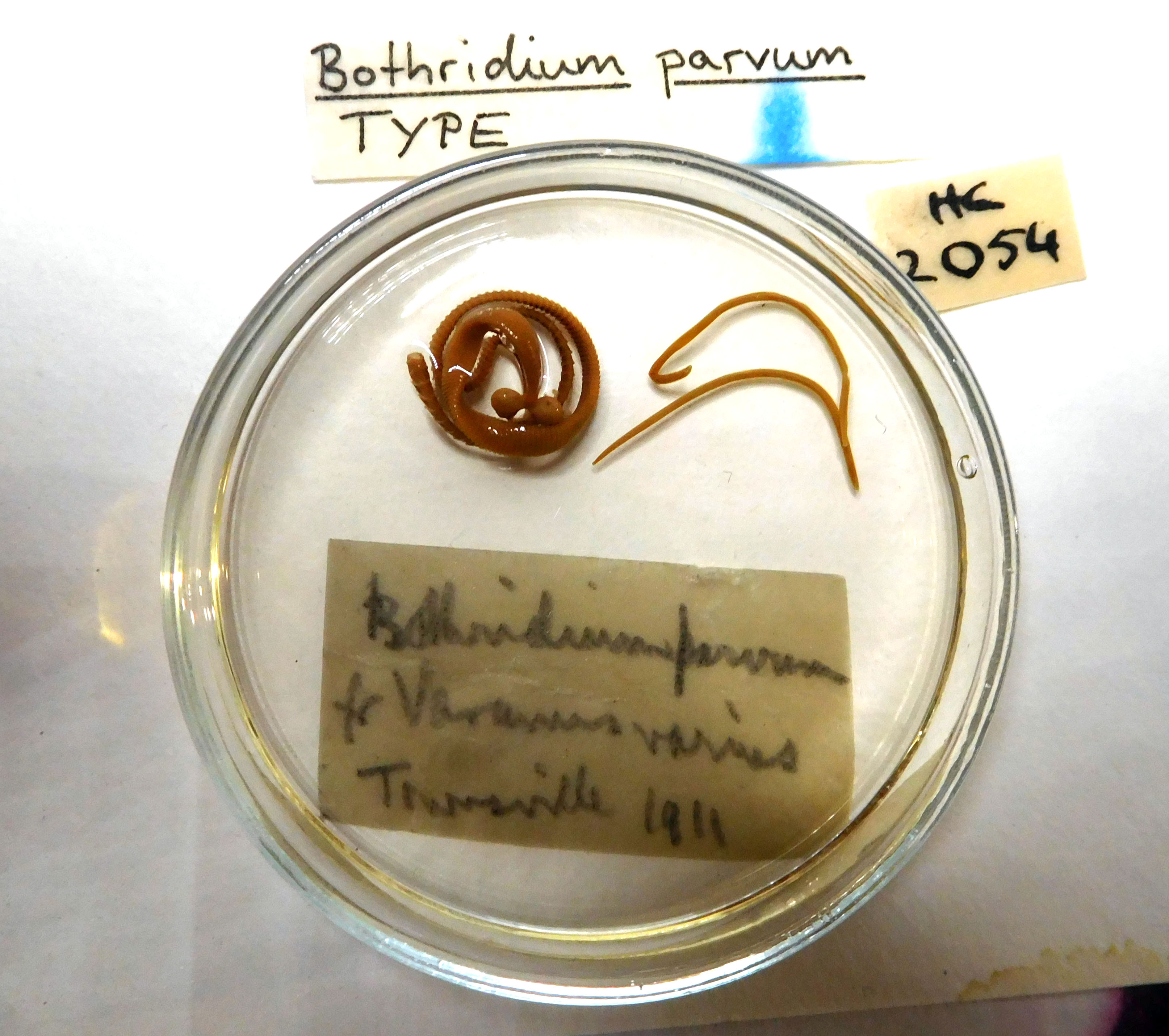 -AHC No. 2054 (holotype)
  -QM No. GL11018 (type) and QM No. GLGL11457 (voucher)

