Line Drawing 1
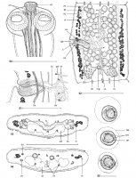
Figs. 5055. Australotaenia grobeli gen. et sp. n. Paratypes BMNH 1968.4.19.1-15,
Figs. 50, 51, 53, 54; Holotype SAM 21402,
Fig. 52. Fig. 50. Scolex, dorsoventral view. Fig. 51. Vagina and cirr... MoreFigs. 5055. Australotaenia grobeli gen. et sp. n. Paratypes BMNH 1968.4.19.1-15,
Figs. 50, 51, 53, 54; Holotype SAM 21402,
Fig. 52. Fig. 50. Scolex, dorsoventral view. Fig. 51. Vagina and cirrus-sac region, ventral view. Fig. 52. Mature proglottis, dorsal (according to de Chambrier 2004, modified). Figs. 53, 54. Cross-sections at the level of the ovary and the posterior part of pregravid proglottis, respectively. Fig. 55. Eggs in distilled water. Abbreviations: ao apical organ; ci cirrus; cs cirrus-sac; do dorsal osmoregulatory canal; em bilayered embryophore; ga genital atrium; lm internal longitudinal musculature; ln longitudinal
nerve; mg Mehlis glands; oe outer envelope; on oncosphere; ov ovary; rm retractor muscles; sc secondary
canal; te testes; ud uterine diverticula; us uterine stem; ut uterus; va vas deferens; vc vaginal canal; vi vitelline follicles; vo - ventral osmoregulatory canal; vs - vaginal sphincter. Scale bars: Figs. 50, 51 = 250 µm; Figs 52-54 = 500 µm; Fig. 55 = 20 µm. |
Line Drawing 2
|
Photo Micrograph
|
Scanning Electron Micrograph
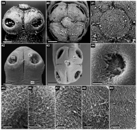
Figs. 3949. Australotaenia grobeli gen. et sp. n. Paratype (BMNH 1968.4.19.1-15). Scanning electron micrographs. Fig. 39. Scolex,
lateral view, note the everted apical organ. Fig. 40. Scolex, apical... MoreFigs. 3949. Australotaenia grobeli gen. et sp. n. Paratype (BMNH 1968.4.19.1-15). Scanning electron micrographs. Fig. 39. Scolex,
lateral view, note the everted apical organ. Fig. 40. Scolex, apical view. Fig. 41. Detail of the apical organ, apical view.
Fig. 42. Scolex, lateral view. Note: small numbers correspond to the figures showing higher magnification images of these surfaces. Fig. 43. Scolex, apical view; apical organ invaginated. Fig. 44. Detail of the apical organ showing long capilliform filitriches, apical view. Fig. 45. Fig. 46. P2@1&&13*".(/&1+"1)%#$(
and coniform spinitriches on the external surface of suckers. Fig. 47.(P2@1&&13*".(/&1+"1)%#$(24,(C&2,12+#($@141+"1)%#$(*4(+%#(.2"ginal
surface of suckers. Fig. 48. P2@1&&13*".(/&1+"1)%#$(24,()*413*".($@141+"1)%#$(*4(+%#(14+#"42&($F"32)#(*3($F)8#"$A(Fig. 49. Capil
&13*".( /&1+"1)%#$( 24,( C&2,12+#( $@141+"1)%#$( *4( +%#( $F"32)#( *3( +%#( @"*&13#"2+1*4( f*4#A( N)2&#( ?2"$V( !1C$A( EU5( TR5( T:5( TE( J( |
 Figs. 5055. Australotaenia grobeli gen. et sp. n. Paratypes BMNH 1968.4.19.1-15,
Figs. 50, 51, 53, 54; Holotype SAM 21402,
Fig. 52. Fig. 50. Scolex, dorsoventral view. Fig. 51. Vagina and cirrus-sac region, ventral view. Fig. 52. Mature proglottis, dorsal (according to de Chambrier 2004, modified). Figs. 53, 54. Cross-sections at the level of the ovary and the posterior part of pregravid proglottis, respectively. Fig. 55. Eggs in distilled water. Abbreviations: ao apical organ; ci cirrus; cs cirrus-sac; do dorsal osmoregulatory canal; em bilayered embryophore; ga genital atrium; lm internal longitudinal musculature; ln longitudinal
nerve; mg Mehlis glands; oe outer envelope; on oncosphere; ov ovary; rm retractor muscles; sc secondary
canal; te testes; ud uterine diverticula; us uterine stem; ut uterus; va vas deferens; vc vaginal canal; vi vitelline follicles; vo - ventral osmoregulatory canal; vs - vaginal sphincter. Scale bars: Figs. 50, 51 = 250 µm; Figs 52-54 = 500 µm; Fig. 55 = 20 µm.
Figs. 5055. Australotaenia grobeli gen. et sp. n. Paratypes BMNH 1968.4.19.1-15,
Figs. 50, 51, 53, 54; Holotype SAM 21402,
Fig. 52. Fig. 50. Scolex, dorsoventral view. Fig. 51. Vagina and cirrus-sac region, ventral view. Fig. 52. Mature proglottis, dorsal (according to de Chambrier 2004, modified). Figs. 53, 54. Cross-sections at the level of the ovary and the posterior part of pregravid proglottis, respectively. Fig. 55. Eggs in distilled water. Abbreviations: ao apical organ; ci cirrus; cs cirrus-sac; do dorsal osmoregulatory canal; em bilayered embryophore; ga genital atrium; lm internal longitudinal musculature; ln longitudinal
nerve; mg Mehlis glands; oe outer envelope; on oncosphere; ov ovary; rm retractor muscles; sc secondary
canal; te testes; ud uterine diverticula; us uterine stem; ut uterus; va vas deferens; vc vaginal canal; vi vitelline follicles; vo - ventral osmoregulatory canal; vs - vaginal sphincter. Scale bars: Figs. 50, 51 = 250 µm; Figs 52-54 = 500 µm; Fig. 55 = 20 µm.  Figs. 3949. Australotaenia grobeli gen. et sp. n. Paratype (BMNH 1968.4.19.1-15). Scanning electron micrographs. Fig. 39. Scolex,
lateral view, note the everted apical organ. Fig. 40. Scolex, apical view. Fig. 41. Detail of the apical organ, apical view.
Fig. 42. Scolex, lateral view. Note: small numbers correspond to the figures showing higher magnification images of these surfaces. Fig. 43. Scolex, apical view; apical organ invaginated. Fig. 44. Detail of the apical organ showing long capilliform filitriches, apical view. Fig. 45. Fig. 46. P2@1&&13*".(/&1+"1)%#$(
and coniform spinitriches on the external surface of suckers. Fig. 47.(P2@1&&13*".(/&1+"1)%#$(24,(C&2,12+#($@141+"1)%#$(*4(+%#(.2"ginal
surface of suckers. Fig. 48. P2@1&&13*".(/&1+"1)%#$(24,()*413*".($@141+"1)%#$(*4(+%#(14+#"42&($F"32)#(*3($F)8#"$A(Fig. 49. Capil
&13*".( /&1+"1)%#$( 24,( C&2,12+#( $@141+"1)%#$( *4( +%#( $F"32)#( *3( +%#( @"*&13#"2+1*4( f*4#A( N)2&#( ?2"$V( !1C$A( EU5( TR5( T:5( TE( J(
Figs. 3949. Australotaenia grobeli gen. et sp. n. Paratype (BMNH 1968.4.19.1-15). Scanning electron micrographs. Fig. 39. Scolex,
lateral view, note the everted apical organ. Fig. 40. Scolex, apical view. Fig. 41. Detail of the apical organ, apical view.
Fig. 42. Scolex, lateral view. Note: small numbers correspond to the figures showing higher magnification images of these surfaces. Fig. 43. Scolex, apical view; apical organ invaginated. Fig. 44. Detail of the apical organ showing long capilliform filitriches, apical view. Fig. 45. Fig. 46. P2@1&&13*".(/&1+"1)%#$(
and coniform spinitriches on the external surface of suckers. Fig. 47.(P2@1&&13*".(/&1+"1)%#$(24,(C&2,12+#($@141+"1)%#$(*4(+%#(.2"ginal
surface of suckers. Fig. 48. P2@1&&13*".(/&1+"1)%#$(24,()*413*".($@141+"1)%#$(*4(+%#(14+#"42&($F"32)#(*3($F)8#"$A(Fig. 49. Capil
&13*".( /&1+"1)%#$( 24,( C&2,12+#( $@141+"1)%#$( *4( +%#( $F"32)#( *3( +%#( @"*&13#"2+1*4( f*4#A( N)2&#( ?2"$V( !1C$A( EU5( TR5( T:5( TE( J( 

