Line Drawing 1
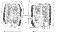
Figs. 25, 26. Vandiermenia beveridgei gen. et sp. n. Paratypes (SAM 32339). Fig. 25. Mature proglottis in dorsal view, uterine stem not figured. Fig. 26. Pregravid proglottis in ventral view. Abbrevia... MoreFigs. 25, 26. Vandiermenia beveridgei gen. et sp. n. Paratypes (SAM 32339). Fig. 25. Mature proglottis in dorsal view, uterine stem not figured. Fig. 26. Pregravid proglottis in ventral view. Abbreviations: cs cirrus-sac; do dorsal osmoregulatory canal; mg Mehlis glands; ov ovary; ud uterine diverticula; va vas deferens; vi vitelline follicles; vo ventral osmoregulatory canal. Scale bars = 500 µm. |
Line Drawing 2
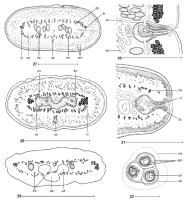
Figs. 2732. Vandiermenia beveridgei gen. et sp. n. Fig. 27. Holotype (SAM 20080), cross-section at the level of the cirrus-sac (redrawn from Johnston 1911). Fig. 28. Paratype (SAM 3778-5), cross-sect... MoreFigs. 2732. Vandiermenia beveridgei gen. et sp. n. Fig. 27. Holotype (SAM 20080), cross-section at the level of the cirrus-sac (redrawn from Johnston 1911). Fig. 28. Paratype (SAM 3778-5), cross-section at the level of the ovary. Fig. 29. Paratype (SAM 3778), cross-section at the anterior part of a pregravid proglottis. Fig. 30. Paratype (SAM 32339), vagina and cirrus-sac
region, ventral view. Fig. 31. Paratype (SAM 32339), cross-section at level of the cirrus-sac.
Fig. 32. Eggs drawn in distilled water, showing the three-layered
embryophore and external small outgrowths. Abbreviations: ci cirrus; cs cirrus-sac; do dorsal osmoregulatory canal; em three-layered embryophore; lm internal longitudinal musculature; ln longitudinal nerve; oe outer envelope; on oncosphere; ov ovary; sm secondary musculature; so small outgrowths; te testes; ud uterine diverticula; us uterine stem; va - vas deferens; vc - vaginal canal; vi - vitelline follicles; vo - ventral osmoregulatory canal. Scale bars: Figs. 27-29 = 500 µm; Figs. 30, 31 = 250 µm; Fig. 32 = 20 µm. |
Photo Micrograph
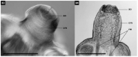
Figs. 33, 34. Vandiermenia beveridgei gen. et sp. n. Paratype (SAM 32339). Photomicrographs. Fig. 33. Pyramidal apex showing the apical part retracted. Fig. 34. Frontal section of the scolex, showing ... MoreFigs. 33, 34. Vandiermenia beveridgei gen. et sp. n. Paratype (SAM 32339). Photomicrographs. Fig. 33. Pyramidal apex showing the apical part retracted. Fig. 34. Frontal section of the scolex, showing the apical organ and the retractor muscle. Abbreviations: ao - apical organ; cm - circular musculature; rm - retractor muscle. Scale bars = 100 µm. |
Scanning Electron Micrograph
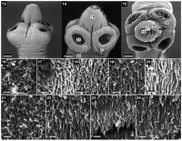
Figs. 1324. Vandiermenia beveridgei gen. et sp. n. Paratypes (SAM 32339). Scanning electron micrographs. Fig. 13. Scolex, lateral view. Fig. 14. Scolex, dorsoventral view. Note: small numbers corresp... MoreFigs. 1324. Vandiermenia beveridgei gen. et sp. n. Paratypes (SAM 32339). Scanning electron micrographs. Fig. 13. Scolex, lateral view. Fig. 14. Scolex, dorsoventral view. Note: small numbers correspond to the figures showing higher magnification images of these surfaces. Fig. 15. Scolex, apical view. Fig. 16. Capilliform filitriches on the centre of the apical organ. Fig. 17. Capilliform filitriches and the gladiate spinitriches on the margin of the apical organ. Fig. 18. Capilliform filitriches and gladiate spinitriches on the upper part of the surface of the pyramidal apex. Fig. 19. Gladiate spinitriches on the middle part of the pyramidal apex surface.
Fig. 20. Gladiate spinitriches on the external upper surface of suckers. Fig. 21. Capilliform filitriches and gladiate spinitriches on the internal surface of suckers. Fig. 22. Capilliform filitriches and gladiate spinitriches on the marginal surface of sucker. Fig. 23. Aristate spinitriches on the surface of the proliferation zone. Fig. 24. Aristate spinitriches on the surface of mature proglottis. Scale bars: Figs. 13-15 = 100 µm; Figs 16-24 = 2 µm. |
 Figs. 25, 26. Vandiermenia beveridgei gen. et sp. n. Paratypes (SAM 32339). Fig. 25. Mature proglottis in dorsal view, uterine stem not figured. Fig. 26. Pregravid proglottis in ventral view. Abbreviations: cs cirrus-sac; do dorsal osmoregulatory canal; mg Mehlis glands; ov ovary; ud uterine diverticula; va vas deferens; vi vitelline follicles; vo ventral osmoregulatory canal. Scale bars = 500 µm.
  Figs. 2732. Vandiermenia beveridgei gen. et sp. n. Fig. 27. Holotype (SAM 20080), cross-section at the level of the cirrus-sac (redrawn from Johnston 1911). Fig. 28. Paratype (SAM 3778-5), cross-section at the level of the ovary. Fig. 29. Paratype (SAM 3778), cross-section at the anterior part of a pregravid proglottis. Fig. 30. Paratype (SAM 32339), vagina and cirrus-sac
region, ventral view. Fig. 31. Paratype (SAM 32339), cross-section at level of the cirrus-sac.
Fig. 32. Eggs drawn in distilled water, showing the three-layered
embryophore and external small outgrowths. Abbreviations: ci cirrus; cs cirrus-sac; do dorsal osmoregulatory canal; em three-layered embryophore; lm internal longitudinal musculature; ln longitudinal nerve; oe outer envelope; on oncosphere; ov ovary; sm secondary musculature; so small outgrowths; te testes; ud uterine diverticula; us uterine stem; va - vas deferens; vc - vaginal canal; vi - vitelline follicles; vo - ventral osmoregulatory canal. Scale bars: Figs. 27-29 = 500 µm; Figs. 30, 31 = 250 µm; Fig. 32 = 20 µm.
Figs. 2732. Vandiermenia beveridgei gen. et sp. n. Fig. 27. Holotype (SAM 20080), cross-section at the level of the cirrus-sac (redrawn from Johnston 1911). Fig. 28. Paratype (SAM 3778-5), cross-section at the level of the ovary. Fig. 29. Paratype (SAM 3778), cross-section at the anterior part of a pregravid proglottis. Fig. 30. Paratype (SAM 32339), vagina and cirrus-sac
region, ventral view. Fig. 31. Paratype (SAM 32339), cross-section at level of the cirrus-sac.
Fig. 32. Eggs drawn in distilled water, showing the three-layered
embryophore and external small outgrowths. Abbreviations: ci cirrus; cs cirrus-sac; do dorsal osmoregulatory canal; em three-layered embryophore; lm internal longitudinal musculature; ln longitudinal nerve; oe outer envelope; on oncosphere; ov ovary; sm secondary musculature; so small outgrowths; te testes; ud uterine diverticula; us uterine stem; va - vas deferens; vc - vaginal canal; vi - vitelline follicles; vo - ventral osmoregulatory canal. Scale bars: Figs. 27-29 = 500 µm; Figs. 30, 31 = 250 µm; Fig. 32 = 20 µm.  Figs. 33, 34. Vandiermenia beveridgei gen. et sp. n. Paratype (SAM 32339). Photomicrographs. Fig. 33. Pyramidal apex showing the apical part retracted. Fig. 34. Frontal section of the scolex, showing the apical organ and the retractor muscle. Abbreviations: ao - apical organ; cm - circular musculature; rm - retractor muscle. Scale bars = 100 µm.
 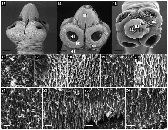 Figs. 1324. Vandiermenia beveridgei gen. et sp. n. Paratypes (SAM 32339). Scanning electron micrographs. Fig. 13. Scolex, lateral view. Fig. 14. Scolex, dorsoventral view. Note: small numbers correspond to the figures showing higher magnification images of these surfaces. Fig. 15. Scolex, apical view. Fig. 16. Capilliform filitriches on the centre of the apical organ. Fig. 17. Capilliform filitriches and the gladiate spinitriches on the margin of the apical organ. Fig. 18. Capilliform filitriches and gladiate spinitriches on the upper part of the surface of the pyramidal apex. Fig. 19. Gladiate spinitriches on the middle part of the pyramidal apex surface.
Fig. 20. Gladiate spinitriches on the external upper surface of suckers. Fig. 21. Capilliform filitriches and gladiate spinitriches on the internal surface of suckers. Fig. 22. Capilliform filitriches and gladiate spinitriches on the marginal surface of sucker. Fig. 23. Aristate spinitriches on the surface of the proliferation zone. Fig. 24. Aristate spinitriches on the surface of mature proglottis. Scale bars: Figs. 13-15 = 100 µm; Figs 16-24 = 2 µm.
Figs. 1324. Vandiermenia beveridgei gen. et sp. n. Paratypes (SAM 32339). Scanning electron micrographs. Fig. 13. Scolex, lateral view. Fig. 14. Scolex, dorsoventral view. Note: small numbers correspond to the figures showing higher magnification images of these surfaces. Fig. 15. Scolex, apical view. Fig. 16. Capilliform filitriches on the centre of the apical organ. Fig. 17. Capilliform filitriches and the gladiate spinitriches on the margin of the apical organ. Fig. 18. Capilliform filitriches and gladiate spinitriches on the upper part of the surface of the pyramidal apex. Fig. 19. Gladiate spinitriches on the middle part of the pyramidal apex surface.
Fig. 20. Gladiate spinitriches on the external upper surface of suckers. Fig. 21. Capilliform filitriches and gladiate spinitriches on the internal surface of suckers. Fig. 22. Capilliform filitriches and gladiate spinitriches on the marginal surface of sucker. Fig. 23. Aristate spinitriches on the surface of the proliferation zone. Fig. 24. Aristate spinitriches on the surface of mature proglottis. Scale bars: Figs. 13-15 = 100 µm; Figs 16-24 = 2 µm. 

