Line Drawing 1
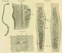
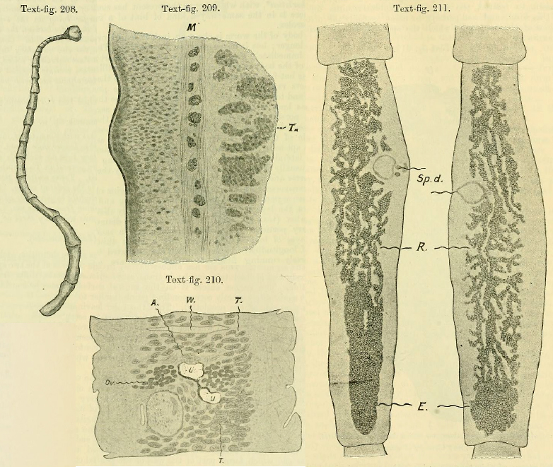
Text-fig. 208. Anoplotaenia dasyuri, enlarged about five times. Text-fig. 209. Anoplotaenia dasyuri, transverse section through part of body-wall. M, two delicate layers of circular fibres between whi... MoreText-fig. 208. Anoplotaenia dasyuri, enlarged about five times. Text-fig. 209. Anoplotaenia dasyuri, transverse section through part of body-wall. M, two delicate layers of circular fibres between which is a special layer of longitudinal fibres grouped into bundles. T, testis. Text-fig. 210. Anoplotaenia dasyuri, longitudinal section of proglottid. A, shell-gland; Ov., ovary, below which is seen the cirrus sac; T., testes; U., uterus, between which and the cirrus sac are seen the coils of the vas deferens cut transversely; W., transverse excretory tube. The posterior part of the proglottid is above. Text-fig. 211. Anoplotaenia dasyuri, two ripe proglottids viewed as transparent objects. E., masses of eggs at posterior end of proglottid; R., reticular portion of uterus; Sp.d., cirrus sac. |
Line Drawing 2
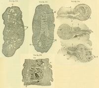
Text-fig. 212. Anoplotaenia dasyuri, transverse section through ripe proglottid. O., ova; U., cavity of uterus. Text-fig. 213. Anloplotaenia dasyuri, transverse section through ripe proglottid. O., ov... MoreText-fig. 212. Anoplotaenia dasyuri, transverse section through ripe proglottid. O., ova; U., cavity of uterus. Text-fig. 213. Anloplotaenia dasyuri, transverse section through ripe proglottid. O., ova, contained in uterus (U.); O1, eggs scattered through parenchyma. Text-fig. 214. Anoplotaenia dasyuri, longitudinal section through proglottid, showing the branched and reticular uterus with ova in smaller and larger clumps. R.S. receptaculum seminis. W., excretory tube. Text-fig. 215. Anoplotaenia dasyuri. Cirrus sac with penis in various stages of retraction. |
Photo Micrograph
|
Scanning Electron Micrograph
|
 Text-fig. 208. Anoplotaenia dasyuri, enlarged about five times. Text-fig. 209. Anoplotaenia dasyuri, transverse section through part of body-wall. M, two delicate layers of circular fibres between which is a special layer of longitudinal fibres grouped into bundles. T, testis. Text-fig. 210. Anoplotaenia dasyuri, longitudinal section of proglottid. A, shell-gland; Ov., ovary, below which is seen the cirrus sac; T., testes; U., uterus, between which and the cirrus sac are seen the coils of the vas deferens cut transversely; W., transverse excretory tube. The posterior part of the proglottid is above. Text-fig. 211. Anoplotaenia dasyuri, two ripe proglottids viewed as transparent objects. E., masses of eggs at posterior end of proglottid; R., reticular portion of uterus; Sp.d., cirrus sac.
 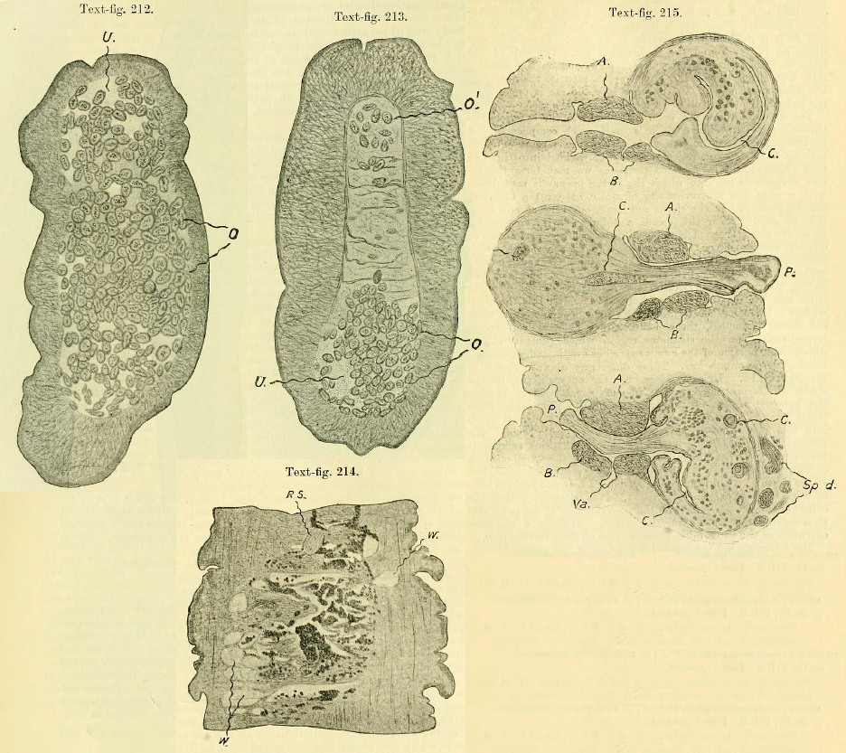 Text-fig. 212. Anoplotaenia dasyuri, transverse section through ripe proglottid. O., ova; U., cavity of uterus. Text-fig. 213. Anloplotaenia dasyuri, transverse section through ripe proglottid. O., ova, contained in uterus (U.); O1, eggs scattered through parenchyma. Text-fig. 214. Anoplotaenia dasyuri, longitudinal section through proglottid, showing the branched and reticular uterus with ova in smaller and larger clumps. R.S. receptaculum seminis. W., excretory tube. Text-fig. 215. Anoplotaenia dasyuri. Cirrus sac with penis in various stages of retraction.

