Cestode Scientific Name
| Species ID | 116 |
|---|---|
| Order | Caryophyllidea |
| Family | Caryophyllaeidae |
| Subfamily | |
| Genus | Atractolytocestus |
| Species | huronensis |
| Authority | Anthony, 1958 |
| Taxonomic Status | Valid |
| Valid Name | |
| Synonyms | |
| Genus Record | No |
| Type Species | Yes |
| Verified | Yes |
| Verified By | T. Scholz |
| Citation(s) |
Anthony, J. D. 1958. Atractolytocestus huronensis n. gen., n. sp. (Cestoda: Lytocestidae) with notes on its morphology . Transactions of the American Microscopical Society 77(4): 383-390. (657) Download PDF |
| Redescription | |
| Scientific Name Notes |
Record Data
| Date (MM/DD/YYYY) | Action | User Name |
|---|---|---|
| 10/13/2005 | Created | Scholz, Hanzelova |
| 10/07/2013 | Modified | |
| 10/03/2016 | Modified | B. Barbeau |
| 04/03/2020 | Modified | T. Scholz |
| 08/17/2021 | Modified | T. Scholz |
Type Host
| Host Class | Actinopterygii | ||||||
|---|---|---|---|---|---|---|---|
| Host Order | Cypriniformes | ||||||
| Host Family | Cyprinidae | ||||||
|
Type Host (Literal) |
|
||||||
|
Type Host (Valid) |
|
||||||
| Additional Host(s) | |||||||
| Site in Host | intestine | ||||||
| Host Notes |
Type Locality
| Country | U.S.A. |
|---|---|
| Body of Water | River Huron |
| Island(s) | |
| City/Region | Ann Arbor, Michigan |
| Coordinates | |
| DD Latitude | |
| DD Longitude | |
| Additional Localities | |
| Locality Notes |
Specimens
| Type Material | |
|---|---|
| Total Number of Type Specimens | |
| Voucher Material | |
| Specimen Notes | Deposition of type specimens not indicated |
Data are given as in original description unless otherwise indicated.

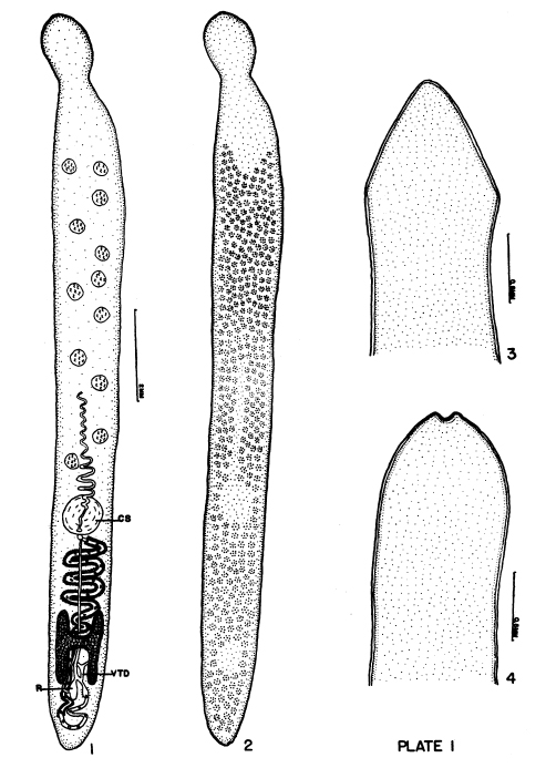 EXPLANATION OF PLATE I.
FIG. 1. Atractolytocestus huronensis in ventral aspect with vitellaria omitted. R-seminal receptacle; VTD-common vitelline duct; CS-cirrus sac. FIG. 2. Same showing the extent of vitellaria. FIG. 3. Scolex of living worm in relaxed condition. FIG. 4. Scolex of a mounted specimen in contracted position.
EXPLANATION OF PLATE I.
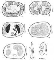
FIG. 1. Atractolytocestus huronensis in ventral aspect with vitellaria omitted. R-seminal receptacle; VTD-common vitelline duct; CS-cirrus sac. FIG. 2. Same showing the extent of vitellaria. FIG. 3. Scolex of living worm in relaxed condition. FIG. 4. Scolex of a mounted specimen in contracted position.  PLATE 2. FIG. 5. Atractolytocestus huronensis. Transverse section through the testes. FIG. 6. Same. Transverse section through the cirrus sac. FIG. 7. Same. Transverse section through the ovarian wings and seminal
receptacle. FIG. 8. Same. Transverse section showing the extent of the post-ovarian vitellaria. FIG. 9. Same. Transverse section through the excretory receptacle. FIG. 10. Same. Embryonic hook. FIG. 11. Same. Egg after eight days of development at room temperature.
PLATE 2. FIG. 5. Atractolytocestus huronensis. Transverse section through the testes. FIG. 6. Same. Transverse section through the cirrus sac. FIG. 7. Same. Transverse section through the ovarian wings and seminal
receptacle. FIG. 8. Same. Transverse section showing the extent of the post-ovarian vitellaria. FIG. 9. Same. Transverse section through the excretory receptacle. FIG. 10. Same. Embryonic hook. FIG. 11. Same. Egg after eight days of development at room temperature. Best viewed in Firefox


