Cestode Scientific Name
| Species ID | 1100 |
|---|---|
| Order | Cyclophyllidea |
| Family | Dilepididae |
| Subfamily | |
| Genus | Eurycestus |
| Species | avoceti |
| Authority | Clark, 1954 |
| Taxonomic Status | Valid |
| Valid Name | |
| Synonyms | |
| Genus Record | No |
| Type Species | Yes |
| Verified | No |
| Verified By | |
| Citation(s) |
Clark, D. 1954. A new cyclophyllidian cestode from the avocet. Journal of Parasitology 40(3): 340-346. (3017) Download PDF |
| Redescription | |
| Scientific Name Notes |
Record Data
| Date (MM/DD/YYYY) | Action | User Name |
|---|---|---|
| 10/03/2005 | Created | Georgiev, Salamatin |
| 04/14/2010 | Modified | |
| 05/11/2016 | Modified | B. Barbeau |
| 10/20/2016 | Modified | A. Phillips |
Type Host
| Host Class | Aves | ||||||
|---|---|---|---|---|---|---|---|
| Host Order | Charadriiformes | ||||||
| Host Family | Recurvirostridae | ||||||
|
Type Host (Literal) |
|
||||||
|
Type Host (Valid) |
|
||||||
| Additional Host(s) | |||||||
| Site in Host | small intestine | ||||||
| Host Notes |
Type Locality
| Country | U.S.A. |
|---|---|
| Body of Water | |
| Island(s) | |
| City/Region | 5 miles N. Lincoln, Nebraska |
| Coordinates | |
| DD Latitude | |
| DD Longitude | |
| Additional Localities | |
| Locality Notes |
Specimens
| Type Material | USNPC No. 49075 (holotype) |
|---|---|
| Total Number of Type Specimens | |
| Voucher Material | |
| Specimen Notes |
Data are given as in original description unless otherwise indicated.


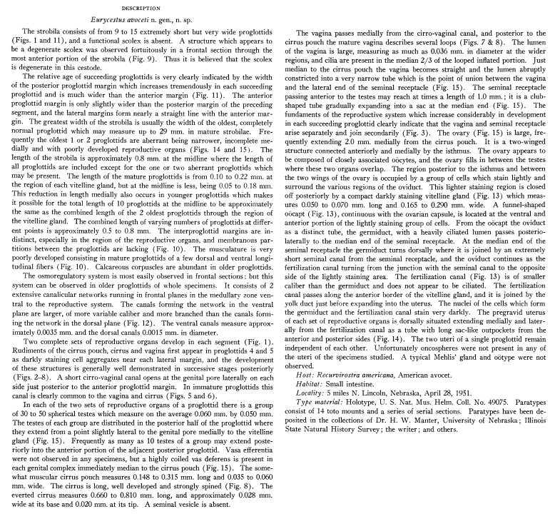
 EXPLANATION OF PLATES All figures are of Eurycestus avoceti n. gen., n. sp. Figures 1 and 11 were drawn with the aid of a projector, and all other figures with the aid of a camera lucida. All scales are in millimeters. PLATE I. FIG. 1. Entire immature worm showing the arrangement of proglottids and the progressive development of the reproductive organs. Protandry is illustrated in this figure. FIGS. 2-8. Various stages in the development of the cirrus pouch, cirrus and vagina; a se- ries of stages usually present in a single strobila. The fundament of the seminal receptacle joins the forming vagina at approximately the stage shown in Fig. 3 where the entire fundament of the seminal receptacle is shown. Testes also appear at approximately this stage and are shown in this figure. Testes are omitted from figures of succeedingly older stages. Fig. 3 was drawn from a frontal section, all others from whole mounts. Figs. 3 to 8 are at the same scale as for Fig. 2. FIG. 9. Frontal section through the most anterior portion of a strobila showing a structure considered to be a degenerate scolex. FIG. 10. Dorsal and ventral longitudinal muscles in three oldest proglottids and lack of membranous partitions between proglottids are shown in a longitudinal section through the median portion of a strobila where reproductive organs are lacking
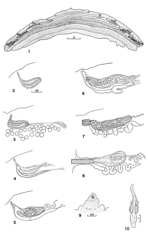
 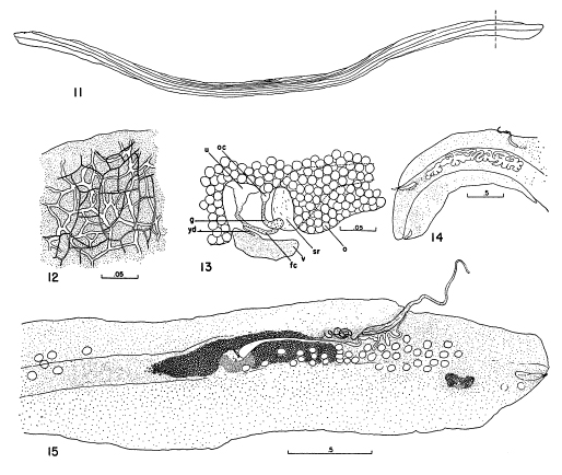 EXPLANATION OF PLATES All figures are of Eurycestus avoceti n. gen., n. sp. Figures 1 and 11 were drawn with the aid of a projector, and all other figures with the aid of a camera lucida. All scales are in millimeters. PLATE II. FIG. 11. Mature strobila in outline showing the arrangement of the proglottids. The mounted specimen measures 29 mm. in width. The portion of the strobila to the right of the vertical line is illustrated in Fig. 15. FIG. 12. Osmoregulatory network in a portion of a frontal section of a mature proglottid. FIG. 13. Diagram of organs in interovarial space, ventral aspect. Lettering: fc, fertilization canal; g, germiduct; o, ovary; oc, o6capt; sr, seminal receptacle; u, uterus; v, vitelline gland; yd, yolk duct. FIG. 14. Pregravid uterus of one side of one proglottid shown in outline. Details of the other reproductive structures omitted. Aberrant posterior proglottid which is frequently in- complete medially is also shown. FIG. 15. Reproductive organs of the portion of the strobila to the right of the line on the strobila illustrated in Fig. 11. Testes are represented by open circles; those to the left of the ovary belong to the adjacent anterior proglottid. The aberrant nature of the oldest proglottid is again indicated by the degenerate reproductive structures in the most posterior proglottid
 Best viewed in Firefox


